Red dirt in the NT


The two weeks I spent in the Northern Territory of Australia were a once-in-a-lifetime opportunity. So many of these moments were new and fascinating.  I was pretty excited to get out of chilly chilly Victoria and get outside into some warm winter weather. As soon as our small plane touched down at Galuwin’ku airport we felt the heat!

20180616_163524

How could you not love this landscape?!

Three of us traveled to the remote Elcho island off the Northern coast of Aus for 2 weeks to be part of a council based Animal Management Program. There is a large amount of community dogs and cats that live in Galuwin’ku (the largest community on Elcho island). The majority of our work was spay and neuter surgeries and distribution of dewormer medication.

IMG_2916

This was one particularly friendly dog that met us on the beach one day and followed us home 🙂 

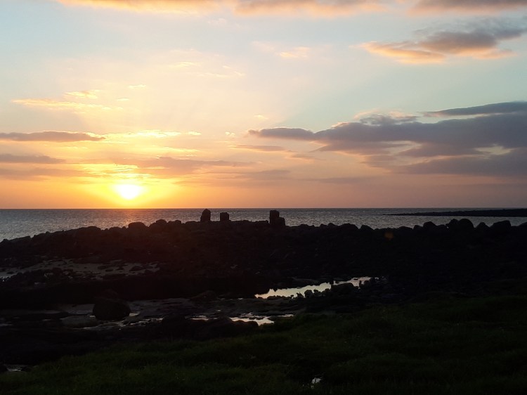
After we first landed we did a little tour of the community and drove past some of the main buildings.  After a few minutes we came across a group of people standing by a dog on the side of the road. He had been hit by a car and was not looking good. We asked if the people wanted us to ‘finish’ him (euthanasia).  In the evening we went to walk by the beach; you can’t go close to the water because it is full of crocodiles. The sunset was amazing. We heard a dog crying and found her laying in a stand of trees in the vines. She was old and riddled with ticks and mites. She belonged to a nearby family who I think moved the dog down to the beach because she had been sick and crying for days. I had never considered how difficult it might be for families to get rid of a dog or euthanize an animal who is sick if there are no vet services around.

20180618_094749

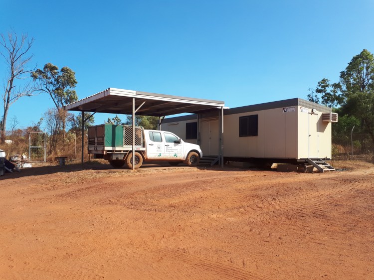
The ‘clinic’ and the ute that we used for all our veterinary work

No one on the island wakes up early; this means that we got to very casually wake up, eat breakfast, and leave the house around 9am… 9:30am…whenever. We often stopped in at the ‘clinic’ (a trailer) and picked up a few things we needed. Then we would drive slowly through the community and stop at family homes and ask them if there was any dogs or cats for ‘operation’ (the term for de-sexing surgery). We would sedate the dogs with their pre-medication drugs at their houses and allow them to get sleepy before loading them into the ute. Children would run and catch the cats for us to put into carriers. At the clinic there was two tables to do surgeries on; you either had to bend over or sit in a chair. One table required you to stack 2 chairs & a pillow on top of each other to be at the right height. There was a desk lamp for added light. We made a makeshift scavenger system for ourselves. The surgical instruments are kept in a saucepan and boiled on the stove each night. Gauze is baked in a canister in the oven. Instruments are scrubbed and sterilized in cold sterile between each surgery. We made swaged on needles as we needed them.  Anesthetic monitoring was done with an SPO2 monitor and our stethoscopes. Each wound was sprayed with Cetrigen purple spray after surgery and dogs received flea spray for a day of relief. All the spays are completed as flank spays: this is because they likely heal faster, if the wound breaks down there is less chance of evisceration, a lot of the patients are either pregnant or lactating and you do not want to disturb the mammary tissue, and its easier to recheck the surgery site from afar. A lot of the animals will remember you and not want to come near you afterwards. Us students got to do almost all of the surgeries by ourselves which was SUCH good experience, and really cool because I got a lot of practice doing the less common flank spays.

IMG_2806.JPG

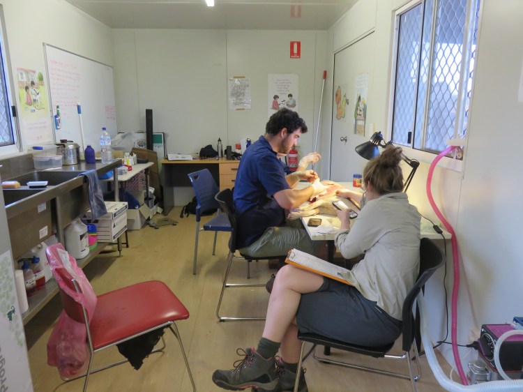
A typical surgical morning.

Occasionally we would do other vet work. We did a couple of new puppy & kitten exams, checked out a lump on a dog, stitched up a dog who had been gorged by a pig, and checked a cat with tail injury.  But, by far, the most thrilling call was—Nigel. Nigel’s family thought he had broken his leg and our whole team was very distressed as we rushed over to his house! Luckily….he must have just stepped on a prickle  and he was totally fine & just as cool as ever.

20180620_143249

Nigel with his mum and his brand new collar!

In Aboriginal communities, especially in a climate like where we were, when someone passes away in the Wet (season) the body cannot be buried because of the humidity and the rain. The body is flown to Darwin where it is held in a morgue until the Dry (season) when it can be returned to the community for a funeral ceremony & burial. Dumbulah is an out station approximately 10 mins from where we were staying. We were invited to watch the funeral ceremony. Funerals can last for days to weeks; they happen for every person who passes away in the community—because of the associated health issues in aboriginal communities, funerals occur fairly often. As we walked closer to the community we could hear singing. When we arrived we saw a few hand painted cloth signs hanging up. The biggest sign was on white cloth, with bright green writing—’ Welcome Home’. Other signs said ‘rest in peace our loving father’, etc. The man’s name was painted.  There was 3 trees as flag poles for the territory flag, the Australian flag, and a third- they all flew at half mast. We sat on the dirt a little ways away and watched some of the singing and dancing. After a short time, a family invited us to come and sit on their mat with them. A lady on our mat explained to us that 2 different clans would sing/dance the next song (I think it was about a sunfish) one after each other. They had differently painted digeridoos.  This was such a valuable life experience for me; I sat there mesmerized for quite a long time.

We had a short meeting with some of the human medical team one morning to talk about a research project that is being run on the island. The medical team is swabbing mothers, children, and infants at certain households to look at skin infections (likely staph/strep). Our job was to take swabs from the dogs who lived at the house as well. The study required swabs from nose, mouth, and perineum.

Most nights we stopped at the beach on the way home to watch the sunset over the water. We would go for walks along the rocks close to the water or sit in a freshwater pool that was a safe distance from the ocean.

20180617_180504

Jess and I woke up early a few mornings to go for a walk on the beach or the road above the ocean to look for crocs or dugongs before breakfast. It was really lovely. We never saw a dugong, but we did see a crocodile!

CDR_0182

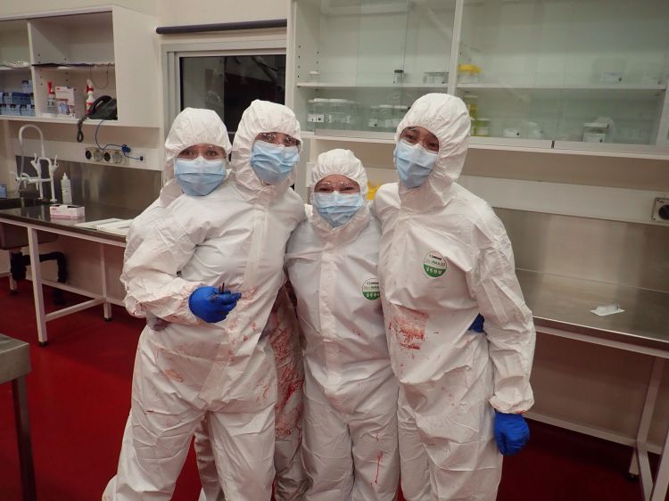
Our fantastic team, we got along swimmingly, and I think all of us grew a bigger spot in our hearts for Aboriginal culture and the NT. 

Farm calls, farmyard surgery, …and chocolate!

I was really looking forward to my Production Animals Rotation and hoping that we would get lots of hands on opportunities with the large animal species that I love working with.

We started the week with a couple of days in the classroom talking through a mastitis case, a reproduction problem on farm, how to get a job in rural practice, etc.

One of the best days during this rotation was when we went to the cattle yards at uni and practiced doing tail vein blood draws, epidurals, inverted L blocks, and paravertebral blocks. The ‘blocks’ are a procedure that the vet usually does prior to surgery where an injection of anesthetic solution is put over nerves & tissues in order to block feeling to that area.  They are a very common procedure in cattle medicine so I am glad we had the opportunity to practice them. We were also able to practice rectal exams & pregnancy diagnosis again. None of the cattle were pregnant.

Wednesday is often everyone’s favourite day. In the morning we visit an abattoir and in the afternoon we go to a chocolate factory. I really enjoyed the abattoir visit; it was a sheep processing facility that produced halal meat. We started at the packing end of the plant where all the cuts of meat in boxes are stored in chilled rooms and packed for shipping. The Australian’s got to experience going into a -20C and -40C freezer. It was entertaining. Then we proceeded up the processing line to where the sheep were stunned and killed. Then we visited the yards outside where the sheep are held when they arrive at the abattoir prior to processing. I really enjoyed this experience because I am interested in food production and a vet’s role in how we are involved in the production of safe, efficient, tasty, humane food.  This is a sensitive topic for a lot of people and I like being educated and involved. In the afternoon we went to the Great Ocean Road Chocolate Factory. It was meant to be a visit to a food processing facility…. I think the university could have picked a better location like a feed mill, or a milk processing plant, but I got free chocolate–so I’m not complaining!  We got to wander around the show room and then went to the back for a special chocolate tasting and spoke with a chocolatier about his techniques and favourite things to create. Some of the chocolate we tried included Australian bush flavors which were really tasty!

20180530_154049

Any day at the chocolate factory is a good day!

We had a fantastic opportunity to do both vasectomies and castrations on rams at the university. In the real world you wouldn’t do both procedures on the same animal because they are required for different reasons. The sheep were anesthetized and resting in a shepherds chair. We worked in partners and were set off to calculate our own drug doses, complete an exam, and get the surgery going. There were other vets around to help us when we got stumped. It was a really fun experience, everything went well and we went back at the end of the day to check on our patients and make sure they were doing fine.

20180531_104153

Jess demonstrating how the shepherds chair works

On one of the days we hopped in a van and drove out a few minutes to a nearby farm to see some sick cows. The first cow had been lame a week ago but looked much better today.  Another heifer looked like she was either walking on her tip toes or dragging them along; after watching her walk around the yards & lifting her feet up we diagnosed her with contracted tendons. She was likely born with them & either they weren’t fixed when she was a calf or they got a lot more significant as she grew older. The third cow we saw was quite skinny & sickly looking. I could FEEL her heart murmur without even using my stethoscope, that’s how impressive it was! She had already been treated a week ago and was not improving, it was decided that she would likely go for post mortem next week if she continued going downhill.

Another day we drove out to a very large sheep farm on a gorgeous property! We stopped and watched someone who was a contracted sheep ultrasounder. He had his own little trailer that he sat in and pregnancy scanned sheep through their flank. His ultrasound probe was different than I have seen before–it had water that sprayed out of it constantly so they he wouldn’t have to waste time by reapplying ultrasound gel. It took him appx 1-2seconds per sheep to determine if she was pregnant and if she was having a single or twin! We all watched completely astonished for a short time. We walked through the woolshed from the 1800s and then spent the afternoon talking about epidemiology cases.

Alpaca farm day! Everyone was pretty excited about this too–because who doesn’t love an alpaca?! We got to practice catching alpacas (basically sneak-attack hugging them around the neck), ultrasounding them for a pregnancy diagnosis, and blood draws. We were also taught the traditional method of getting alpaca’s to sit down–I forget the proper word! You can tie their legs up underneath them and then they will sit calmly for you to perform a procedure or transport them. There was also a few males that needed to be castrated so we got to ‘share an alpaca’ and practiced our farmyard castrations.

This was a fun rotation and it makes me excited for some placements I have booked with large animal practices back home!

20180607_153209

On the last day we had to give presentations—This is my group mates who brought a model cow all the way from the shed into the seminar room just for a demonstration! 

X-rays. Buffalo. Microscopes. Whale.

The diagnostics rotation is very different from everything else we’ve done this year. We were back to sitting in chairs and focusing on one topic all day long–it was a difficult transition for some of us. This was not my favorite rotation, but it was very good to practice reading blood smears, blood results, x-rays, and performing post mortem exams. The rotation was split into three components: clinical pathology, radiology & imaging, and anatomic pathology.

Clinical Pathology

This rotation was mostly self directed learning. We had a variety of cases to work through ourselves. We had several hematology and biochemistry cases to look at (blood work) and cytology cases (slides of cells).  We spent many hours looking down a microscope and compiling our answers all together.  One morning we all gathered in the lab and practiced doing packed cell volumes (PCVs) and blood smears. We also practiced doing urinalysis and looking at the sediments for the cells under the microscope.  The ‘sediment’ in a urine sample is all the cells and crystals and other things present in the urine; you can collect them on a microscope slide after you spin the sample in a centrifuge machine.

20180524_100712

Blood under the microscope

Radiology & Imaging

The second component of our diagnostics rotation was radiology and imaging. We started with the musculoskeletal system, and then thorax and abdomen. We were given multiple x-ray films from cats, dogs, and horses. We had normal x-rays to look at as well.  There were several radiology cases to work through and then we had to present a couple cases and our findings to the radiologist. When animals in the hospital needed a x-ray our group would go and assist with positioning the patient on the table and exposing the x-ray. We also looked at the images with the specialists in the radiology room. On the final day of the rotation we had a ‘positioning assessment’ where we had to position a stuffed dog model for different radiographic views.

20180525_134757

My group in the CT room practicing the positioning of ‘Emily’ for her x-rays

I have always enjoyed ultrasounds, so I thought it was very cool when we got some hands on ultrasound practice during this rotation. The first time we got to practice was on the ‘phantom’ which is a specially made box with spheres and wires hidden inside gel that we used the probe to detect. The phantom helps us learn how to manipulate the settings on the ultrasound to better find an image.  The next day we had a LIVE dog to practice on!  We started by finding the kidney in a longitudinal plane and then moving the ultrasound probe to image it in a different view, then moving the probe around the abdomen to look at the spleen, liver, intestines, and bladder. We were supposed to have a 3rd day to practice with the ultrasound and a live patient, but there was no dog available to us. We ended up ultrasounding each other—I am happy to report that I have a gall bladder but possibly only 1 kidney (jk I probably have two kidneys but we couldn’t find the other one).

Anatomic Pathology

Post mortems can be completed on animals as a diagnostic test to help determine the cause of death–especially if it was unable to be determined while the animal was still alive. The first post mortem we did was a congo buffalo from the free range zoo.  Before she died she was walking very weirdly; Her movements looked similar to a disease we see in horses called ‘Stringhalt’. She was immobilized with Etorphine prior to euthanasia. Etorphine is a drug that is very dangerous to humans and can be absorbed through skin and cause immediate cardiac arrest (depending on dose). Because of this we had to wear full PPE (personal protective equipment) and be briefed on what symptoms to watch out for in ourselves and our colleagues. We had someone on standby with a reversal drug. As well, we marked off the leg that the buffalo was darted in & removed that leg prior to starting our necropsy.

congo buffalo

A Congo Buffalo! (No, I also had no idea what they looked like before we saw her)

The second post mortem we performed was a dog that had died due to a dog attack. It was very interesting to see the full extent of the damage from the bite wounds that were not visible externally. The damage likely wouldn’t have been able to be identified with any imaging modalities either.  This experience will influence how I treat future trauma and dog attack cases because I am now better able to understand how many ‘hidden problems’ these patients have.

Our 3rd and most exciting post mortem was a whale! My group was very lucky to be involved in this post mortem.  The whale was stranded in shallow water and was unable to be resuscitated.  Our group and our pathology professors took a little field trip down to the zoo to perform the necropsy at their facilities. We wore full PPE again to protect ourselves against diseases that we might pick up from a marine mammal. Our team was organized—one person was in charge of measurements, ensuring all appropriate samples were collected & organized, others were in charge of examining different body systems. We discovered a massive gastric impaction with sea grass. When we opened one of the stomach compartments there was a large sheet of plastic as well. After doing some research for our post mortem report, we learned that this species of whale eats squid, not sea grass. We suspect that after eating the plastic she may been more inclined to eat an abnormal diet and started eating the sea grass because she had abdominal pain. Unfortunately, she was also pregnant.

32512068_10155161941376394_6060239459276816384_n

Hanging out in our PPE suits! 

My favourite part of this rotation was the post mortems, especially because we got to work with some very interesting species. However, I’m excited to move on to the next one and get more action in my day!


“To Cut is to Cure”

We had a 1 week surgery rotation at the university hospital. The university is a referral center with specialist surgeries being performed so we didn’t get to do much ourselves. We scrubbed into as many surgeries as possible and held tissues or passed instruments or operated the suction for the surgeon.  We worked long days; coming in early each day to check on surgery patients from the day before. We also called patients a couple days after their surgeries to check on them and see how they were doing post-op. We wrote discharge notes and surgical reports. Surgical reports are different than a normal medical history. We made sure to include all the instruments used, the type of scalpel and suture, the pattern of closures, exact dimensions of any incisions, and carefully explained each procedure. Each evening we had rounds where we talked about the surgical cases for the day and looked at the pre- and post-operative imaging (x-rays, or CT scans or MRIs).

One of my first patients this week was a beautiful dog who had a significantly sized mass removed from his soft palate (roof of his mouth). Once the surgeon took the mass out there was a very large fistula which needed to be repaired with a skin graft. I was reading some of the articles the surgeon had with him to assist the procedure. It was very complicated, and the anatomy went over my head…that’s the thing about the field of veterinary medicine & surgery… there is always so much more to learn and do and practice! He stayed in hospital for a few days and had to be stomach tubed his meals (who wants to eat when you have a skin graft in your mouth anyways?!). When we went out for walks I had to stop him from trying to pick up sticks with his mouth! Unfortunately, after a couple days one side of his skin graft had failed and he needed a repeat surgery to close the fistula in the roof of his mouth again. After freshening the edges and making another mini-graft the fistula was closed. As far as I know, this patient recovered really well after his revision surgery!

I watched a couple of other mass removal surgeries this week, they were not quite as cool as the soft palate resection + graft. One poor dog had a mass in her rectum that was very uncomfortable. Another dog was very prone to developing lumps all over her body and needed those removed!

One girl in my group watched four splenectomies or something crazy like that this week. I managed to watch one!  The university has this very cool surgical tool which makes splenectomies a lot easier; it basically cauterizes all the vessels and stops all the bleeding so the surgery is much faster and cleaner.


One day there was a cria (baby alpaca) that had come in for surgery. He was too big to stay in the small animal hospital so he stayed with his mum in the horse barn and then came to our small animal surgery team to have his bony sequestrum removed from his leg.  A sequestrum is basically a dead piece of bone sitting within the limb; they can be associated with infections and sometimes draining tracts will form. The CT scan of his leg was particularly impressive!

If any spays or neuters were scheduled my group members and I were meant to do them. I watched one of my friends do a spay and a couple of my other friends did castrations. I was very unlucky all week and my cases kept cancelling or no-showing on me! I didn’t get to do any surgeries this week.


Towards the end of the rotation I had a very complicated surgical case! The patient was in the ICU.  I completed my rotation in the ICU earlier this year. The ICU team determined that the patient had a septic abdomen. Which meant that he had a bacterial infection in his abdomen—this is really not ideal! We learned that this dog had previously been diagnosed with severe hydrocephalus and had previous brain surgery to put in a ventriculoperitoneal shunt (VP shunt). Very simply —this is a tube that goes from the brain (and tracks under the skin) to the abdomen where it drains the cerebrospinal fluid that builds up in the brain. The excess fluid cannot be in the brain and if it is drained to the abdomen it can be reabsorbed into the body there.

Image result for dog with hydrocephalus

Please enjoy this Google image of a puppy with hydrocephalus. His head is abnormally enlarged and domed. http://www.dogscatspets.org/hydrocephalus-in-dogs/hydrocephalus-dogs/

Unfortunately, the presence of this shunt complicated matters. We didn’t know if the bacterial infection in his abdomen had started in his brain or if it has started in his abdomen and then had tracked up to his brain. The surgery team spoke with the ICU team and then the surgery team liaised with the neurology team. Eventually we decided that it was in our patient’s best interests to complete his abdominal surgery first and then bring in the neurology team to remove his shunt. Then, if he recovered well in ICU he would require another brain surgery in a couple weeks to replace the VP shunt in his brain (this patient cannot live without the shunt). Both surgeries went really well! I helped put in the esophageal feeding tube at the end of surgery.  I’ve been trying to keep up on how this patient was doing by asking my friends who were on their ECC rotation in the ICU room.