Yay and neigh!

Not sure if that blog title makes sense… but its horse related!

I have always wanted to work with large animals after graduation, I have always had the hardest time finding equine experience. So one of my goals for final year was to gain more confidence working with horses so that after graduation I could be a mixed animal veterinarian who was willing to see horses and enjoyed the calls.  I had a 3 week placement at an equine clinic, there were boarded veterinarians on staff and they saw horses with many different kinds of problems.

For the first two weeks I was working with the surgery team. The mornings we spent in the surgical ward and then often the afternoons were filled with lameness exams.  My role often involved helping ‘drop’ (sedate) the horse in the padded recovery room and then move it onto the surgery table with the help of a winch. Here is a quick video showing how a horse is moved into surgery.  I could then help scrub the surgical site (for a really long time!!!) to make sure the area was clean before the surgeon made his incision. On the first day I scrubbed into a cryptorchid surgery and tried my hand at some suturing. I still haven’t been given the chance to do a gelding (castrating a male horse) myself and I cannot wait until I have the chance to do one. I watched a couple of arthroscopic surgeries; such as 3rd carpal bone fracture repairs on racehorses. One of the more interesting surgeries I got to watch (twice actually!) was a mandibular tooth retropulsion. The mandibular teeth are in the back of the jaw, on the bottom, so if they need to be extracted they are very hard to pull out the traditional way. It is much easier to drill a hole in the bottom of the jaw and then retropulse (basically hammer) the tooth out with a peg. This surgery required a lot of skull radiographs to ensure that the surgeon was drilling and hammering the peg in the right direction in order to remove the correct tooth. I was in charge of taking the radiographs and this is probably why these were my favourite surgeries to be involved with.

20180717_132423

Just in case you ever wondered how big a horse’s tooth is, here you go!

I also helped complete the treatments for all of the hospitalized horses. This meant giving injections, checking bandages, flushing intravenous catheters, taking horses for walks to graze, cold hosing wounds, etc.  In one part of the hospital there is a large model horse, we put it in front of the stall of any horses who are lonely or anxious without a friend –often it helps them calm down! One patient I helped with had gotten a large stick rammed up into his groin, another had extremely sore feet as he had a condition called laminitis.

20180722_201819

The lonely horse friend; conveniently on wheels!

During the first week there was an Ag Tour going on in the county; the tour group stopped by the practice on two different days. We had 3 stations in the hospital to explain to the people on the tour what actually goes on inside a big equine referral hospital. One of the tour groups actually got to watch through the windows of the surgical suite as we completed one of the mandibular tooth retropulsion surgeries.

On some afternoons we would load up the truck and go out to see horses at stables or farms. We often went to stables and looked at horses training for different competitions, we did lameness exams and joint injections. I often helped scrub the site prior to the injections. I did my first nerve block (abaxial) on this placement!  When owners are preparing to take their horses across the border into the United States they need to be checked by a veterinarian and have a recent blood test (Coggins) completed before they are allowed to enter the states. We did quite a few of these exams and blood tests. Often the paperwork for these horses can be monotonous but there is now a new app that allows vets to fill out information and take photo records on their phones! I also watched quite a few euthanasias and on a couple of horses we completed post mortems to find out why they had died. The intern and I got to practice finding our landmarks and performing joint injections.

20180721_161606

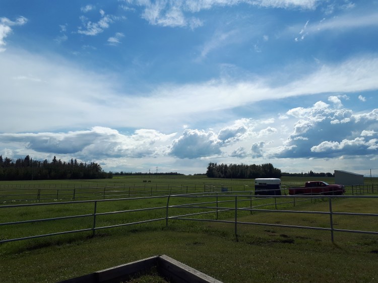
One of the lovely summer views we had while out on call. At this particular location we were dealing with a horse that refused to get onto the trailer!

I got to use some of the practice-owned horses to hone my lunging (an important skill for conducting lameness exams and just being a generally horse-competent person in general), reproductive rectal exam with the ultrasound, and placing a jugular IV catheter.  I also practiced putting in a nasogastric tube; it is an easy enough procedure to do when there is a calm situation.  NGT’ing is an important diagnostic for working up a colic case and I’m nervous for the first time I’ll have to get a NGT into a colicking horse (which can often be very stressful!).

On the third week I worked in the reproduction barn. It was the end of the season and appointments were slowing down–but this was good because it left me lots of time to ask questions. I spent a lot of time running out to mares in pens and giving them injections of hormones to help manage their reproductive cycles so that we could get them bred (so they could have a foal next year).  When we weren’t giving injections to mares we often had them inside the barn for a reproductive tract ultrasound exam; we would look at the body of the uterus, the horns, the ovaries and any follicles or corpus lutea that were present. If a horse had already been bred then we would monitor her for fluid build up (that could ruin a pregnancy) and preg-check her at 14 days to see if she had twins or not, and then assess the fetus’s heartbeat at 25 days. Later in gestation, the owners could bring their horse back if they wanted to know the foal’s gender (filly vs colt). I also got to watch a couple of stallion collections using an artificial vagina— if you wanna talk about dangerous jobs; collecting stallions is definitely one!

There was a cute little loft apartment on site that ended up being really handy–I stayed there when I did on call with the vet. Some of our emergency calls included: a miniature horse foaling, colicking horses, a broken skull, a horse who wouldn’t get on a trailer, and wounds. Other notably cool appointments included a horse enucleation, a horse with a bullet in his leg, a suspected Strangles case, and a suspected Potomac Horse Fever case!

I had a great time on this placement, I really enjoyed the chats with vets in the vehicle, bonding with the intern, the variety of appointments to see, and the friendly nature of everyone at the clinic! Thanks a million Delaney Veterinary Services 🙂

P.S: This was the only placement that gave ME a thank you card at the end of my time there, I was so blown away by this!!


Hay gurl

My trip home over Christmas break was so refreshing. It was just what I needed before heading back to Australia for the final year of my degree. The day I landed back in Melbourne it was 39C. It was a bit of a shock to the system…and naturally being me–I couldn’t wait to get to the beach.

20180128_202224

How is this only 20 mins from my house?

The next morning we started our Equine Internal Rotation at the horse hospital on campus.  This was the rotation that everyone said was difficult, you just have to get through it, and people cry every year. I’m not sure if I took the same rotation or not… but my group had the best time! The whole class is divided up into groups and we spend the  year with those same people as we go through our university rotations.

Whenever a new patient would be admitted to the hospital or come in for a day procedure one of us students had to claim the patient and we would work with that case for the duration of the hospital stay. This also meant that we had to know all the diagnostic tests, findings, and treatments that were run on the patient–we presented everything at the morning and afternoon rounds.

My first case was very interesting. The foal had been admitted to the hospital the day previously with difficulty breathing. It had a tracheostomy tube put in his trachea so he could breath. When I first came in the foal started showing some neurological signs so we decided to investigate further. X-rays showed us that the foal had actually broken it’s neck. This foal needed to be euthanized. I did it; I place very high value on these experiences during vet school.

We call something a ‘textbook case’ when it presents with all the exact signs and symptoms of a disease that you would read about in a textbook. We had a textbook ‘Recurrent Airway Obstruction’ (RAO) case.  It basically means the horse has allergies. It is an important and fairly common equine disease and something that I should likely know well for my board exam! I was pleased that this case came in during my rotation so I could see it in real life and not just read about it. We did a test called a bronchoalveolar lavage (BAL) on the horse which includes putting fluid into the horse’s lungs and then sucking it back up to get a sample of the cells present. Depending on what cells we find this can tell us information about the disease the horse has.

Last year I volunteered in the equine hospital as a 3rd year student. One of the patients that was very memorable came back in for a bit of a check up. This horse is a particularly interesting case because he has a collapsed trachea. This is typically something we see more commonly in dogs and definitely not in horses! His collapsed trachea has already been repaired and he is living comfortably with 2 stents in it to make sure it stays open and he can breathe!

Over the course of the 2 weeks we had a couple of very sick patients–one of them was in isolation. This means we had to start super early, put on all our protective wear and go into his isolation stall to check on him.

Snapchat-1695992760

Luckily there is a cafe in town that opens at 5am. We head there for some real caffeine. 

One of the things I really wanted to see during this rotation was surgery. I specifically was interested in seeing a colic surgery–unfortunately that didn’t happen. But I did get to watch an extremely beautiful Clydesdale undergo an impressive surgical procedure. She had bilateral hock arthroscopy procedures and an ocular cyst removal done. Horse surgery to me is so much more impressive than surgery on cats and dogs. Horses need to be anesthetized and then picked up on a huge winch/crane system so that they can be placed on the surgical table. It can be dangerous to anesthetize horses because of their large size—it causes problems with their lungs! When we first knocked this horse out there was about 13 people running around who all had jobs to prepare the horse for surgery—putting in a urinary catheter, preparing, and scrubbing the surgical sites, applying monitoring equipment, hooking up anesthesia tubes and drugs, positioning the horse, etc etc. Usually I love jumping in and getting involved but this was one time I was happy to be a fly on the wall and just observe all the action around me! It felt like a dream!

Orthopedic work is a big deal in horse medicine. We saw a lot of lame horses during this rotation.  No, a ‘lame horse’ is not a horse we don’t like, it means a horse that is painful in its legs or feet and is not walking normally. This means we watch the horse walk, trot, canter, etc. We check to see if the horse is more lame on a hard surface or a soft surface or while turning or at different speeds. Using a variety of techniques and tests we try to figure out exactly where in the leg the horse is painful! Nerve blocks are a common test we do; we inject a little bit of anesthetic into specific nerves in the leg. This means the horse loses sensation to the area of the leg that that nerve goes to. If we ask the horse to trot and the lameness is gone–then we have a better idea of the location in the leg that the horse is painful!  We can also use x-ray, ultrasound, nuclear scintigraphy, and MRI to help.

Horses are great at injuring themselves! They are impressive and beautiful animals but they are amazing at getting into tricky situations. My next patient was a horse who had been kicked. The wound on his leg went right into the joint space. This can be very dangerous if bacteria are able to grow and then spread around the body. Using a small needle you can stick it directly into the joint and take a sample of his joint fluid. We also flushed out the joint with saline and then left some liquid antibiotics in there before bandaging it all up! We monitored him closely to make sure the infection was healing and not spreading.


A couple of my group’s favorite patients were there for almost the whole 2 weeks of our rotation. One of them was an older teaching horse with a medley of problems but the sweetest personality! We made sure to spend a few extra minutes with her every day. The other horse we loved was a very small stallion who presented with only one eye!  He was very sweet. During his physical exam every day he let me hug him while my partner did the exam.

Snapchat-781925505_LI (2)

A very scientific sample of the discharge taken from the eyeless horse’s socket. We needed to make sure it wasn’t infected. 

Most days we were working 11 or 12 hour days. One day we managed to actually get out of the horse hospital at a decent time and went to the beach right afterwards because it was a nice day. Just as we were getting ready for bed my cell phone rang and we were out the door and back to the hospital for a colicking horse!  Colic is a confusing term–it basically means anything that can give a horse abdominal pain.  An initial colic work up is providing pain medication, rectal palpation (to feel the abdominal organs), checking vital signs, nasogastric tubing, and abdominal ultrasound.  We did all of these tests because we were trying to figure out if the horse needed to go to surgery or if we could treat him medically.  The horse was quite sick but his clinical signs weren’t making sense with any of the more common causes of colic. We decided it was probably best to monitor the horse over night and reassess him the next morning.  During our exam the next day we noticed that he hadn’t urinated since arriving at the hospital and being on IV fluids over night.  I passed a urinary catheter as far as I could up his urethra–until I hit a blockage. Then we put a scope (which is a tiny camera) up his urethra until we saw a stone!! That’s why he couldn’t pee. We did something called a ‘belly tap’ which where we insert a needle into the horse’s abdomen to get a sample of fluid—we discovered that it was urine! This horse had a very rare condition. A stone (we call them uroliths) had formed in his urine and traveled down his urethra until it got stuck and could not move. The horse could not pee and this kept going on until his bladder burst and his abdomen filled up with urine (uroabdomen). Unfortunately, this is very unusual in an adult horse and this horse likely needed at least 2 surgeries to have a chance of recovering—his owner elected to euthanize him. We did the post mortem the next day and the pathology we found definitely confirmed that that was the right decision for this horse.

The next evening we got a referral for a very sick pregnant mare coming in. My whole group decided to stay after hours even though we weren’t all on call.  This horse had hepatic lipidosis –which is a liver disease and was very sick. We were concerned that she was going to abort her foal or go into labor much too early. We decided that she needed to be induced to foal. We prepared all our equipment and debriefed for CPR on the foal.  We were all so tired from being on call and working long days–none of us had food for dinner. We made a call to a kebab shop for sustenance.

We spent a lot of time with that mare and didn’t leave until close to midnight. We had case presentations to give the next day—and we weren’t finished preparing yet! Me and my partner picked the uroabdomen horse to present on because it is such an interesting case! We got home, and finished our presentation and made cupcakes for our last day as a thank you to the equine hospital team! And yes, we modeled them after our patients….

20180209_084539

The cupcakes were modeled after our favourite teaching horse!

Extra big thank you to all the vets, residents, and nurses who put in an effort to teach us, answer our questions, and help us along these past two weeks!