KEEP CALM OR I WILL BOLUS THE PROPOFOL

Image result for keep calm or i will bolus the propofol

Propofol is a very commonly used drug in the veterinary world. We use it to help patients fall asleep before surgery.

My last rotation of the year was spent in the anesthesia department at the university hospital. This was a rotation that my whole group wished had been scheduled earlier in the year. We were taught lots of valuable information about drugs that would have been very helpful during our placements–but better late than never!

The anesthesia department at the hospital runs almost all of the anesthetic protocols for any procedure going on in the hospital–so this means cats, dogs, horses, and occasionally other fun animals!  An anesthetic protocol is required for any patient that needs to be asleep for a procedure– like taking an x-ray or having a surgery done.

Each student was required to be ‘primary’ on a case for the day and read their patient’s file and then develop an anesthetic plan that would appropriately match the patient’s diseases and procedure. This is complicated. Especially when your patient has multiple diseases and you can’t give them certain drugs.

20181031_113313

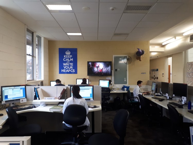
Jessica reading up on her patient’s medical history. There is a TV in our student lounge connected to one of the surgical suites. On this day, I sat & ate my lunch and watched a thoracotomy. 

After doing a physical exam on our patients, creating an anesthetic protocol, and preparing all our equipment and machines we would draw up our drugs into syringes. The first step is then administering a pre-medication drug to the patient to make them a little sleepy. After that we would collect our patient from the wards, bring them to the prep room and place an IV catheter in their vein. We also placed an endotracheal tube (so we can control their breathing), and finished prepping them for their surgery or procedure. During surgery we monitor the patient’s vital signs to make sure their heart is beating properly, they are breathing correctly, and other important things. Once the surgery is finished we are in charge of making sure the patient wakes up smoothly and safely.

One day I monitored an anesthetic for 7 hours, one of my wonderful group mates brought me food from home and the rest of the team tagged me out for a break 🙂

During some of the afternoons we had tutorials where we learned about different stages of the anesthetic protocols and could ask any questions we needed to.

We all had to take complete charge of a patient (with no help from supervisors and nurses) and we were assessed to make sure we could do everything appropriately and safely. My patient was in hospital for a toe amputation. Everything went smoothly!

Some of the patients that I created anesthetic protocols and monitored for were getting CT scans, MRIs, spays, foreign body removal surgeries, etc.

Unfortunately, this post can’t be too much longer without talking about a lot of fancy drug names and anesthetic equipment. I found it helpful to practice using new drugs and talk about protocols with the experts!  At the end of the 2 weeks…we were done! But actually, DONE! No more rotations 🙂 next step…. graduation!

45261786_1928843004077418_8720174372830052352_n

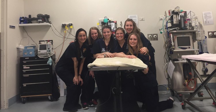
Last day of DVM4! We are in the prep-room in front of some of the anesthetic machines we had been using during our rotation. 


Farm calls, farmyard surgery, …and chocolate!

I was really looking forward to my Production Animals Rotation and hoping that we would get lots of hands on opportunities with the large animal species that I love working with.

We started the week with a couple of days in the classroom talking through a mastitis case, a reproduction problem on farm, how to get a job in rural practice, etc.

One of the best days during this rotation was when we went to the cattle yards at uni and practiced doing tail vein blood draws, epidurals, inverted L blocks, and paravertebral blocks. The ‘blocks’ are a procedure that the vet usually does prior to surgery where an injection of anesthetic solution is put over nerves & tissues in order to block feeling to that area.  They are a very common procedure in cattle medicine so I am glad we had the opportunity to practice them. We were also able to practice rectal exams & pregnancy diagnosis again. None of the cattle were pregnant.

Wednesday is often everyone’s favourite day. In the morning we visit an abattoir and in the afternoon we go to a chocolate factory. I really enjoyed the abattoir visit; it was a sheep processing facility that produced halal meat. We started at the packing end of the plant where all the cuts of meat in boxes are stored in chilled rooms and packed for shipping. The Australian’s got to experience going into a -20C and -40C freezer. It was entertaining. Then we proceeded up the processing line to where the sheep were stunned and killed. Then we visited the yards outside where the sheep are held when they arrive at the abattoir prior to processing. I really enjoyed this experience because I am interested in food production and a vet’s role in how we are involved in the production of safe, efficient, tasty, humane food.  This is a sensitive topic for a lot of people and I like being educated and involved. In the afternoon we went to the Great Ocean Road Chocolate Factory. It was meant to be a visit to a food processing facility…. I think the university could have picked a better location like a feed mill, or a milk processing plant, but I got free chocolate–so I’m not complaining!  We got to wander around the show room and then went to the back for a special chocolate tasting and spoke with a chocolatier about his techniques and favourite things to create. Some of the chocolate we tried included Australian bush flavors which were really tasty!

20180530_154049

Any day at the chocolate factory is a good day!

We had a fantastic opportunity to do both vasectomies and castrations on rams at the university. In the real world you wouldn’t do both procedures on the same animal because they are required for different reasons. The sheep were anesthetized and resting in a shepherds chair. We worked in partners and were set off to calculate our own drug doses, complete an exam, and get the surgery going. There were other vets around to help us when we got stumped. It was a really fun experience, everything went well and we went back at the end of the day to check on our patients and make sure they were doing fine.

20180531_104153

Jess demonstrating how the shepherds chair works

On one of the days we hopped in a van and drove out a few minutes to a nearby farm to see some sick cows. The first cow had been lame a week ago but looked much better today.  Another heifer looked like she was either walking on her tip toes or dragging them along; after watching her walk around the yards & lifting her feet up we diagnosed her with contracted tendons. She was likely born with them & either they weren’t fixed when she was a calf or they got a lot more significant as she grew older. The third cow we saw was quite skinny & sickly looking. I could FEEL her heart murmur without even using my stethoscope, that’s how impressive it was! She had already been treated a week ago and was not improving, it was decided that she would likely go for post mortem next week if she continued going downhill.

Another day we drove out to a very large sheep farm on a gorgeous property! We stopped and watched someone who was a contracted sheep ultrasounder. He had his own little trailer that he sat in and pregnancy scanned sheep through their flank. His ultrasound probe was different than I have seen before–it had water that sprayed out of it constantly so they he wouldn’t have to waste time by reapplying ultrasound gel. It took him appx 1-2seconds per sheep to determine if she was pregnant and if she was having a single or twin! We all watched completely astonished for a short time. We walked through the woolshed from the 1800s and then spent the afternoon talking about epidemiology cases.

Alpaca farm day! Everyone was pretty excited about this too–because who doesn’t love an alpaca?! We got to practice catching alpacas (basically sneak-attack hugging them around the neck), ultrasounding them for a pregnancy diagnosis, and blood draws. We were also taught the traditional method of getting alpaca’s to sit down–I forget the proper word! You can tie their legs up underneath them and then they will sit calmly for you to perform a procedure or transport them. There was also a few males that needed to be castrated so we got to ‘share an alpaca’ and practiced our farmyard castrations.

This was a fun rotation and it makes me excited for some placements I have booked with large animal practices back home!

20180607_153209

On the last day we had to give presentations—This is my group mates who brought a model cow all the way from the shed into the seminar room just for a demonstration! 

“To Cut is to Cure”

We had a 1 week surgery rotation at the university hospital. The university is a referral center with specialist surgeries being performed so we didn’t get to do much ourselves. We scrubbed into as many surgeries as possible and held tissues or passed instruments or operated the suction for the surgeon.  We worked long days; coming in early each day to check on surgery patients from the day before. We also called patients a couple days after their surgeries to check on them and see how they were doing post-op. We wrote discharge notes and surgical reports. Surgical reports are different than a normal medical history. We made sure to include all the instruments used, the type of scalpel and suture, the pattern of closures, exact dimensions of any incisions, and carefully explained each procedure. Each evening we had rounds where we talked about the surgical cases for the day and looked at the pre- and post-operative imaging (x-rays, or CT scans or MRIs).

One of my first patients this week was a beautiful dog who had a significantly sized mass removed from his soft palate (roof of his mouth). Once the surgeon took the mass out there was a very large fistula which needed to be repaired with a skin graft. I was reading some of the articles the surgeon had with him to assist the procedure. It was very complicated, and the anatomy went over my head…that’s the thing about the field of veterinary medicine & surgery… there is always so much more to learn and do and practice! He stayed in hospital for a few days and had to be stomach tubed his meals (who wants to eat when you have a skin graft in your mouth anyways?!). When we went out for walks I had to stop him from trying to pick up sticks with his mouth! Unfortunately, after a couple days one side of his skin graft had failed and he needed a repeat surgery to close the fistula in the roof of his mouth again. After freshening the edges and making another mini-graft the fistula was closed. As far as I know, this patient recovered really well after his revision surgery!

I watched a couple of other mass removal surgeries this week, they were not quite as cool as the soft palate resection + graft. One poor dog had a mass in her rectum that was very uncomfortable. Another dog was very prone to developing lumps all over her body and needed those removed!

One girl in my group watched four splenectomies or something crazy like that this week. I managed to watch one!  The university has this very cool surgical tool which makes splenectomies a lot easier; it basically cauterizes all the vessels and stops all the bleeding so the surgery is much faster and cleaner.


One day there was a cria (baby alpaca) that had come in for surgery. He was too big to stay in the small animal hospital so he stayed with his mum in the horse barn and then came to our small animal surgery team to have his bony sequestrum removed from his leg.  A sequestrum is basically a dead piece of bone sitting within the limb; they can be associated with infections and sometimes draining tracts will form. The CT scan of his leg was particularly impressive!

If any spays or neuters were scheduled my group members and I were meant to do them. I watched one of my friends do a spay and a couple of my other friends did castrations. I was very unlucky all week and my cases kept cancelling or no-showing on me! I didn’t get to do any surgeries this week.


Towards the end of the rotation I had a very complicated surgical case! The patient was in the ICU.  I completed my rotation in the ICU earlier this year. The ICU team determined that the patient had a septic abdomen. Which meant that he had a bacterial infection in his abdomen—this is really not ideal! We learned that this dog had previously been diagnosed with severe hydrocephalus and had previous brain surgery to put in a ventriculoperitoneal shunt (VP shunt). Very simply —this is a tube that goes from the brain (and tracks under the skin) to the abdomen where it drains the cerebrospinal fluid that builds up in the brain. The excess fluid cannot be in the brain and if it is drained to the abdomen it can be reabsorbed into the body there.

Image result for dog with hydrocephalus

Please enjoy this Google image of a puppy with hydrocephalus. His head is abnormally enlarged and domed. http://www.dogscatspets.org/hydrocephalus-in-dogs/hydrocephalus-dogs/

Unfortunately, the presence of this shunt complicated matters. We didn’t know if the bacterial infection in his abdomen had started in his brain or if it has started in his abdomen and then had tracked up to his brain. The surgery team spoke with the ICU team and then the surgery team liaised with the neurology team. Eventually we decided that it was in our patient’s best interests to complete his abdominal surgery first and then bring in the neurology team to remove his shunt. Then, if he recovered well in ICU he would require another brain surgery in a couple weeks to replace the VP shunt in his brain (this patient cannot live without the shunt). Both surgeries went really well! I helped put in the esophageal feeding tube at the end of surgery.  I’ve been trying to keep up on how this patient was doing by asking my friends who were on their ECC rotation in the ICU room.

Eyeballs, ovaries, and anesthesia!

Its finally the start of our Easter break and its given me a chance to write another post. This has been a full on semester…the material is a lot more clinically relevant and interesting. I love that we get more hands on experience. This is the fun stuff, the stuff that counts, its what I’ve been looking forward to for years.

Recently I had a really good week of practical classes:

The first one was our Ophthalmology Practical class.  I’ve helped out with tons of eye examinations at work over the years, but only got to use the equipment myself a few times. I have never been overly interested in eye cases but this class was a lot of fun for me. We had our ophthalmology lectures a few days earlier which were full of different pictures and explanations of what to look for. Our lovely teaching greyhounds were present and we got to practice Schirmer Tear Tests (STT (measures tear production)), fluorescein eye stains (highlights wounds or ulcers in the eye), and examination of the eyeball with a focal light and an optivisor. I’m starting a clinical placement at a small animal clinic this week and I am really hoping to get a chance to practice my examination skills.

We had our second Bovine Reproduction Practical class where we practiced rectal palpation on female cattle. Our main objective was to attempt to find and palpate the cervix, bifurcation of the uterus, and both ovaries. I was able to find all the structures; one of our cows even had a cystic ovary. That ovary was a lot larger and easier to find than the normal healthy ovaries. The week before we had an Equine Reproductive Practical class where we felt for the same structures in horses. It was good practice to go between species and think about the differences in anatomy. Horse ovaries are bigger and you feel for them higher up than cow ovaries.

6tag_170317-170054

We both found ovaries! Well on our way to diagnosing pregnancy…. 

Our Small Animal Reproductive Practical class finished off our clinical skills in our reproduction block. Unfortunately there was no dogs or cats present in our class so we mostly used microscopes to examine slides of swab samples. Cytology (the examination of cells) is one of the best ways to determine which stage of a cycle the bitch (female dog) is in. It is very important to know what stage she is in so that she can be bred on the right day and get pregnant with puppies. Looking through microscopes is definitely not my favorite part of vet med but after this class I understand a lot more what to look for in these cases.

My favorite practical class that week was our Equine Anesthesia class. We were split into 2 groups of students. Half of us were given a drug protocol and had to calculate drug doses and draw up our medications. The other half of the class had to complete a physical exam on our patient. My roommate and I were in the same group and we felt quite confident about how to start and complete a physical exam on a horse because of our time volunteering in the horse hospital. After we completed our exam we had to clean, prep, and insert a jugular catheter so we could administer the premedication drugs. Our group had a ‘Triple Drip’ drug protocol which is very common in horses. Then we proceeded to anesthetize our horse with our maintenance drugs. When the patient was asleep we were responsible for monitoring his vital signs and recording everything on the anesthetic record. We also practiced intubation. I’ve done intubations in cats and dogs before, horses are different because it is a ‘blind intubation’. That means that you cannot see exactly where you are placing the tube and instead have to rely fully on feel and knowledge of anatomy.

6tag_290317-164459

I am holding the maintenance drugs we used for our TIVA (total intravenous anesthesia) Triple Drip protocol and equine intubation tubes. 

So vet school? Loving it!

After a full 9 weeks of class I’m ready for a 2 week break to catch up on lectures and complete some clinical placements…