“To Cut is to Cure”

We had a 1 week surgery rotation at the university hospital. The university is a referral center with specialist surgeries being performed so we didn’t get to do much ourselves. We scrubbed into as many surgeries as possible and held tissues or passed instruments or operated the suction for the surgeon.  We worked long days; coming in early each day to check on surgery patients from the day before. We also called patients a couple days after their surgeries to check on them and see how they were doing post-op. We wrote discharge notes and surgical reports. Surgical reports are different than a normal medical history. We made sure to include all the instruments used, the type of scalpel and suture, the pattern of closures, exact dimensions of any incisions, and carefully explained each procedure. Each evening we had rounds where we talked about the surgical cases for the day and looked at the pre- and post-operative imaging (x-rays, or CT scans or MRIs).

One of my first patients this week was a beautiful dog who had a significantly sized mass removed from his soft palate (roof of his mouth). Once the surgeon took the mass out there was a very large fistula which needed to be repaired with a skin graft. I was reading some of the articles the surgeon had with him to assist the procedure. It was very complicated, and the anatomy went over my head…that’s the thing about the field of veterinary medicine & surgery… there is always so much more to learn and do and practice! He stayed in hospital for a few days and had to be stomach tubed his meals (who wants to eat when you have a skin graft in your mouth anyways?!). When we went out for walks I had to stop him from trying to pick up sticks with his mouth! Unfortunately, after a couple days one side of his skin graft had failed and he needed a repeat surgery to close the fistula in the roof of his mouth again. After freshening the edges and making another mini-graft the fistula was closed. As far as I know, this patient recovered really well after his revision surgery!

I watched a couple of other mass removal surgeries this week, they were not quite as cool as the soft palate resection + graft. One poor dog had a mass in her rectum that was very uncomfortable. Another dog was very prone to developing lumps all over her body and needed those removed!

One girl in my group watched four splenectomies or something crazy like that this week. I managed to watch one!  The university has this very cool surgical tool which makes splenectomies a lot easier; it basically cauterizes all the vessels and stops all the bleeding so the surgery is much faster and cleaner.


One day there was a cria (baby alpaca) that had come in for surgery. He was too big to stay in the small animal hospital so he stayed with his mum in the horse barn and then came to our small animal surgery team to have his bony sequestrum removed from his leg.  A sequestrum is basically a dead piece of bone sitting within the limb; they can be associated with infections and sometimes draining tracts will form. The CT scan of his leg was particularly impressive!

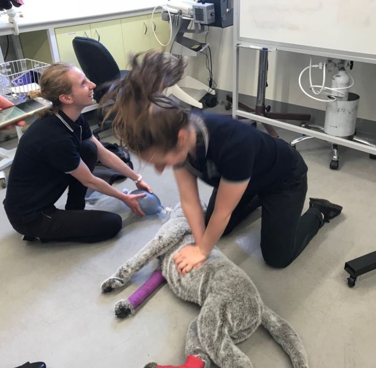
If any spays or neuters were scheduled my group members and I were meant to do them. I watched one of my friends do a spay and a couple of my other friends did castrations. I was very unlucky all week and my cases kept cancelling or no-showing on me! I didn’t get to do any surgeries this week.


Towards the end of the rotation I had a very complicated surgical case! The patient was in the ICU.  I completed my rotation in the ICU earlier this year. The ICU team determined that the patient had a septic abdomen. Which meant that he had a bacterial infection in his abdomen—this is really not ideal! We learned that this dog had previously been diagnosed with severe hydrocephalus and had previous brain surgery to put in a ventriculoperitoneal shunt (VP shunt). Very simply —this is a tube that goes from the brain (and tracks under the skin) to the abdomen where it drains the cerebrospinal fluid that builds up in the brain. The excess fluid cannot be in the brain and if it is drained to the abdomen it can be reabsorbed into the body there.

Image result for dog with hydrocephalus

Please enjoy this Google image of a puppy with hydrocephalus. His head is abnormally enlarged and domed. http://www.dogscatspets.org/hydrocephalus-in-dogs/hydrocephalus-dogs/

Unfortunately, the presence of this shunt complicated matters. We didn’t know if the bacterial infection in his abdomen had started in his brain or if it has started in his abdomen and then had tracked up to his brain. The surgery team spoke with the ICU team and then the surgery team liaised with the neurology team. Eventually we decided that it was in our patient’s best interests to complete his abdominal surgery first and then bring in the neurology team to remove his shunt. Then, if he recovered well in ICU he would require another brain surgery in a couple weeks to replace the VP shunt in his brain (this patient cannot live without the shunt). Both surgeries went really well! I helped put in the esophageal feeding tube at the end of surgery.  I’ve been trying to keep up on how this patient was doing by asking my friends who were on their ECC rotation in the ICU room.

The First Cat and Dog Rotation!

I feel like there is a significant lack of cat and dog jokes around to make into a quirky title for this blog…

After my group finished our equine rotations we needed to switch our brains into ‘smallies’ mode because we had 3 weeks of Emergency and Critical Care (ECC) and Small Animal Medicine rotation ahead of us.

The Intensive Care Unit (ICU) room is located in the middle of the university hospital and all the most sick or critical patients get transferred there. Often the team is so busy they don’t leave the room and have no idea whats going on in the other departments!  We either worked day shifts or evening shifts–which were the ones I preferred.  This meant that after normal day time hours most of the other hospital departments had gone home–so if a patient needed a special procedure or diagnostic test we did it ourselves instead of transferring to another department.

Again, as a student we were meant to take a case and be “in-charge” of the treatment and monitoring of that animal while it was in hospital. If an animal presented to the hospital it was called over the loudspeaker and we went to the reception area to triage the patient and consult with the owners.

The first patient I saw ended up being the most complicated case we had all week! The cat was initially suspected to have hepatopathy (a fancy way of saying liver disease) and renal (kidney) disease. After further treatment and diagnostics we discovered both an intussusception and a gastric foreign body with bi-cavitary effusions, hypothermia, hypovolemia, and hypotension…which is a real fancy way of saying that this cat was very very sick!  Each day we had rounds where we could sit outside in the gardens and discuss emergency topics. This patient was a great patient to initiate discussions on identification of shock and management of critically ill patients.

On our next shift we had a blocked cat, which is something I had seen a lot of at my previous job! It is not an un-common problem in male cats.  My housemate went to a conference last year and listened to a lecture about sacroiliac blocks (epidurals) for cats during this procedure. We use epidurals quite commonly in cattle but less so in our small animal patients. One night we were lucky enough to have a cadaver cat to practice epidurals on as well as other emergency skills like jugular catheters, tracheotomies, thoracocentesis, and urethral catheterization.

Another evening, there must have been something in the water in Werribee because we had multiple patients present after they feasted on the laundry room products!! One dog ate everything in site—detergent, soap, wine, bleach, smashed glass, etc. One cat ate lily laundry detergent and lilies are particularity toxic to cats!!

In Australia it is quite common to see snake bite cases! The university is currently working on SnakeMap which is this cool project that collects GPS coordinates of where snake bites occur which will help veterinarians manage the cases more efficiently. We had a few patients present to us in different stages of the course of the disease. It was fun being involved in these cases and I will miss them when I practice in Canada one day.

In the true spirit of emergency we had a couple cases which were rushed directly to the ICU room.  My group stood back in awe and watched the well-oiled ECC team perform CPR on a ferret who was involved in a dog attack.  Within minutes we had experts from the anesthesia team and the exotic specialist vet at the ICU room to assist with the uncommon patient.  Another patient in the ICU needed a pericardiocentesis preformed.   This is a procedure where the vet will stick a special needle through the body wall into the sac surrounding the heart (without poking the heart) to remove fluid—often blood–which is preventing the patient from breathing and pumping blood around their body properly.  This is a really cool technique because it is both diagnostic & therapeutic. This means that by performing a pericardiocentesis we can often get a diagnosis of the disease we are dealing with and we can also TREAT that condition at the same time.

WhatsApp Image 2018-04-22 at 10.06.35 PM

Practicing CPR on stuffed animals during our tutorial. But with that hair-do…. I’m not sure if its CPR or headbanging 😛

My group then transitioned into the Small Animal Medicine Department for a slower paced and more detailed rotation. We had tutorials on nutrition for cats and dogs and discussed diets required for different medical conditions and how to best formulate that into a treatment management plan. We also talked about antibiotics and prudent use.  Lastly, we had a CPR tutorial where we practiced our technique on life-sized stuffed animal patients.  We had a ton of fun practicing because we searched up YouTube and played “Stayin’ Alive” on loud to help us keep on time (compressions should be done at a tempo of 120 beats/min)!

I was in charge of another critically ill patient this week that was transferred back and forth from Medicine, ECC, and Surgery. This cat initially presented with blood in his urine. However, he was very anemic as well. Over the course of the week he ended up needing at least 2 blood transfusions. At one point we were considering giving him dog blood (xenotransfusion) because we didn’t have any feline donors.  Personally, I didn’t know that you could even do that without an animal dying and was completely mind blown! The cat continued to get sicker during the week as we tried to figure out an explanation for his many problems. We performed x-rays, ultrasounds, multiple blood tests, and even an exploratory laparotomy (surgery).  This cat had a very guarded prognosis and I was amazed and delighted at the end of the week when he was pacing around the ward and meowing at me for more food in his bowl (and not through his stomach tube!).

Wednesday was cardiology day! We shadowed the specialist cardiologist that comes to the university. My group sat in on all his morning appointments to watch echocardiograms (an ultrasound of the heart) and ECGs. I had a patient that had come in earlier in the week for something unrelated and when I did my physical exam I heard an abnormally low heart rate, a few tests later and an appointment booked in with the cardiologist and we had diagnosed her with a serious heart condition requiring surgical implantation of a pacemaker.

20180313_114208

Sometimes you need several ECG pages to read in order to diagnose the heart condition your patient has…

And on Thursday we had extra training with the specialist oncologist. She was really lovely and spent some time helping us to better feel lymph nodes in dogs and cats. This is something that I have always struggled to find in normal patients who do not have enlarged nodes.  Several of the patients I saw this week were cancer patients in various stages of diagnosis, staging, and treatment.  I now have a better understanding of chemotherapy drugs, what to do when you think you may have found cancer in a patient, how to treat and diagnose cancer.

These 3 weeks provided tons of opportunities for reviewing multiple different diseases and we had a lovely time in the hospital!