Adventures of a Mixed Practice Vet (Student)

I’ve just completed my very last placement of final year! I still have rotations to do (they are organized by the university not myself) before I’m totally finished. I booked my placement at this mixed animal vet clinic to be 1 month long. I wanted more time to get to know the vets, the area, and follow up on patients—which are features that you do not get when your placements are shorter.

I started at 9am on Monday, by 9:30am the first patient was in the hydraulic squeeze in the bovine exam room and I was in charge of replacing his rectal prolapse. Luckily the prolapse was quite small and I really only needed to put in a couple stitches.  As for other cattle work —I tried to jump on all these calls because this was the hands on experience that I wanted.  I went to a lot of herd health appointments– usually the first appointments of the day.  This is usually an appointment that occurs every 2 weeks at a dairy farm to assess the health & pregnancy status of the herd.  I preferred to get my practice with rectal palpation and trying to diagnose cows as ‘pregnant’ or ‘open’.  Some cool things other than pregnancies that I felt included cystic ovaries and a mummified fetus from an aborted pregnancy. Sometimes I watched the ultrasound screen while the vet checked the cows. You can tell how far along the pregnancy is by looking at the size of the embryo on the screen.

One particularly memorable call was helping out a heifer who having difficulty calving. She had already been going several hours and the farmer said her calf was dead. By the time we got out to the farm the rain was bucketing down from the dark clouds. We pulled up to an uncovered head gate with the heifer waiting for us.  With thunder and lightening rolling in we got to work. Calving calls usually requires a lot of lube…. and I mean a lot. I dumped a 1L bucket of lube into a pail and used a stomach pump and a hose to pump all the lube into the birthing canal (sorry for that mental image all you non-vet people). We also slathered both our arms in lube and got to work.  The dead calf came out piece by piece. I felt like we really helped that heifer; and bonus: we didn’t get struck by lightening!

We did a left displaced abomasum (LDA) surgery in clinic. I explained what this was in one of my previous blog posts about a cattle placement. Working on our large animal calls in clinic is often much nicer than on farm because all of our equipment is right there, we are in a controlled (& heated!) environment and we can often be a lot cleaner/more sterile than in the field. The LDA surgery was performed similar to others I have seen except this time we were able to wear gowns, gloves, and perform a proper scrub in the sink before starting.

We saw quite a few cattle with joint infections. Often all this takes is a small cut or wound over the incision that allows bacteria to come in and colonize. Sometimes these patients can be treated and sometimes they cannot. Close to the end of my placement a calf with a very badly infected joint came in. The situation looked grim but the farmer was game to try and save the calf.  We sedated the calf until it was laying on the ground. I was in charge of clipping the hair and scrubbing the foot (imagine how long it takes to clean a cow’s leg that has never been bathed ever in its life!) before we tapped the joint (stuck a needle into the joint space). We ended up draining a lot of the STINKY infected joint fluid before we flushed out the joint space and injected some antibiotics directly into the space. I applied a big bandage to the cows leg and was chatting to the farmer—it was almost 10 minutes later I realized I had chunks of joint material & dirt hanging off my face. #classy.

We drove out one day to an extremely nice property to see 3 highlander calves ((the fluffy ones!) who are apparently famous on instagram?!) that were coughing.  We suspect that they had lungworm because they seemed healthy otherwise and started coughing whenever they had to run around. On our way back to the clinic one of the tires on the truck blew off… so that was quite the adventure!

20180807_202747

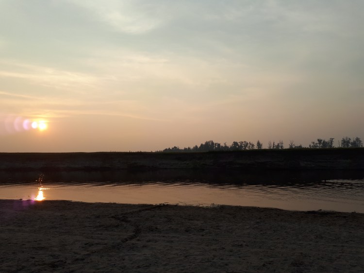
I went for a sunset swim in the river with friends after work

Seeing ‘bull with broken penis’ on the appointment scheduler is always interesting. I think we saw about 3 of these cases while I was at this practice. One of them just had a bad infection and the other bull had a massive abscess all surrounding his sheath. I drained out & cleaned the abscess. Both the bulls went on antibiotics and they will be out for the rest of the breeding season this year.

Someone brought in a much older cow and an unrelated calf who both had walking issues. On rectal exam of the older cow you could feel spondylosis (extra bone growth) between all her vertebrae.  This is an age related change and was probably the bone just trying to stabilize itself. We didn’t think she could be safely bred any longer.

We also saw lots of sick cows which allowed me to practice my physical exams. We even had a few conversations about dart guns, treating wild cattle, and which drugs were the best to use because of amount, effectiveness, and depth of penetration of the dart (subcutaneous administration vs intramuscular). And yes, then I went and checked out a dart gun & some of the 10ml darts that are being used.

There was always something going on with the cats and dogs in the clinic. I placed drains in a couple of different patients with wounds. The drains go into ‘dead space’ beneath the skin of the animal and assist in draining out fluid that would delay or prevent healing. The first drain I placed was in a dog who had a lumpectomy surgery and his incision had dehisced. He came back in a week or so later for a suture check and then for his sutures to be removed; the wound had healed up great! On my last day of placement a large breed dog who had been attacked by another had a head & neck FULL of wounds. I placed 2 drains in his neck. A few days after I finished my placement one of the vets texted me pictures of his healing wounds!

20180901_170144.jpg

I had only been at the practice 4 days… and I already had clients leaving messages for me 🙂 

Speaking of wounds, because of the rural area there was a lot more appointments of pets that had been fighting, were attacked, etc. I looked at many wound patients, or hit-by-car patients, watched enucleation surgeries, and pulled porcupine quills out of dogs (one of my favorite appointment types)! I even saw my first cat with quills stuck in her face — cats usually run away so this was interesting.

During my first week there was a young puppy who refused to walk. The x-rays showed a broken leg. I got to see this puppy several times over the next 4 weeks as he came back in for bandage changes.

There was one dog who was diagnosed with bladder cancer, this is typically a fast moving and impossible to cure cancer. We were trying to manage this patient to be as comfortable as possible. She was having issues urinating so I helped to pass an indwelling urinary catheter that would sit in her bladder and allow her to pee. I also helped pass a urinary catheter in a male cat who was blocked (usually a stone stuck in his urethra preventing him from urinating).

One day a dog came in for ‘excessive slobbering’. This problem actually turned out to be a lot more interesting when we discovered the dog was unable to close his mouth and likely had a nerve paralysis!

snapchat-865248869.jpg

We had to do an emergency surgery on a dog who we thought swallowed a sock or a rope. It turned out that this dog was just obsessively eating grass until he packed his stomach solid with it. 

I was able to perform castration and spay surgeries by myself. As well, I taught the veterinarians & techs about a new surgical technique we learned (the Millers knot) that is really good for crushing tissue. The vets loved it and are going to use it themselves now! I scrubbed in and helped in a very large breed dog spay & a pregnant spay and even a c-section on a Frenchie! The vet let me deliver the last puppy myself— slipperier than expected!  I had a super interesting conversation with a vet one day because we had several puppy vasectomies and hysterectomies booked in. These are not common de-sexing procedures because they leave the testicles & ovaries behind in the patient which can continue to produce hormones. There are some breeders in the area that were requesting these surgeries. Speaking of puppies & breeders…. we did several first puppy exams. This means that I stood in a room of 5-10 puppies and got to cuddle them all!… I mean… examine… and then give them needles (vaccines). I’m talking white Labs, chocolate Labs, Beagle crosses, German shepherds….

We saw a young mastiff dog was was limping heavily, after some x-rays we discovered a severly diseased elbow with a condition called Ununited Anconeal Process (UAP) which requires a specialist surgery.

We did a couple of post mortems on rabbits! Not your typical case….

I started all of the small animal consults by myself, did my own exam, took samples, etc. before chatting to the vet about my suspected diagnosis. Then we went back in and finished the appointments together. I saw a puppy who was only a few weeks old that was dripping fluid from his nose & sneezing everywhere. We suspected pneumonia, although neither me or the vet had ever seen this condition in an animal so young.  We saw an old Caviler King Charles Spaniel with an extremely impressive display of decompensated heart disease with a murmur that likely could have been heard in the next room. We sedated a dog who’s owner thought he had porcupine quills stuck in his ears. Once I examined his ear canals I found a grass awn and managed to pull it out for him! Imagine just how irritating that would be?!

Snapchat-57809322

Feelin’ good! 

THEN, there was all the horse calls! We did a couple of pregnancy checks on mares which was good to do in a different environment because I did a lot of this at my last placement. One of the vets at this practice had also done another course on chiropractic & acupuncture treatment for animals so she had several appointments (horses & dogs) come in for this.

I got to try my hand at floating teeth with a power float… harder than it looks… and I still haven’t mastered the ability to see all the teeth at the back of the horse’s mouth without climbing inside…

On another day we were on our way to see a horse that was acting neurological, by the time we arrived the horse had died. We spoke with some people on the farm and then took a blood sample for testing; we suspected West Nile Virus. When the results came back a week or so later that was not the case but a different disease that could have been prevented by vaccination was found instead!

We went to another farm to euthanize a very old horse. And another farm to check on a couple of horses with wounds. One horse had a wound high up in his armpit, another had a wound on the back of his leg with his tendons exposed! Back at the clinic we saw some miniature horses that had been attacked by dogs and had significant wounds. It was an after-hours call that took us almost 2 hours to clean up & stitch back together. I heard that they are doing well.

The smoke had been so bad in the area (from forest fires in the next province) for almost 2 weeks that I wasn’t surprised when we went and did an exam on 2 horses with respiratory issues from the terrible air quality.

There was a horse with a really bad case of mastitis. Usually this is a problem that we see more commonly in cattle & if you see it in a horse it is easily treated. That was not the case for this horse. When we saw her her udder was very swollen, firm, and quite painful. We checked on her almost daily & prescribed her a whole slew of medications.  I also put in a couple of intra-mammary antibiotic treatments & an IV jugular catheter. Treating her with intra-mammaries reminded me that a horse udder has a different anatomy than a cow’s udder. One day we got a call that now her udder was looking better but she was 3-legged lame & couldn’t walk! Confused, we drove out to the farm again. The horse was hungry but didn’t want to move to the grass. Her legs and tendon sheaths were all swollen. We suspected she may have had a drug reaction & pulled her off all her medication. The next day she seemed to be on the up & up!

So that’s a lot of fun stories! And I didn’t even mention all the fun I got to have in the evenings & weekends being home with my friends/fam…. coffee dates, farm visits & parties, dinner with my grandmas, learning to ride a motorbike & pull a trailer, markets & fairs, swimming, campfires, drinks….

I really enjoyed myself at this practice. The vets treated me like a doctor, like a colleague, and I felt my opinion was valued and appreciated. Thanks a million to BarrNorth Veterinary Services and I wish you a fantastic fall and winter season ahead!

Resized_20180822_124321_3959

The clinic had a taco-in-a-bag customer appreciation day so my mom and a family friend came for a tour of the practice! 

Hay gurl

My trip home over Christmas break was so refreshing. It was just what I needed before heading back to Australia for the final year of my degree. The day I landed back in Melbourne it was 39C. It was a bit of a shock to the system…and naturally being me–I couldn’t wait to get to the beach.

20180128_202224

How is this only 20 mins from my house?

The next morning we started our Equine Internal Rotation at the horse hospital on campus.  This was the rotation that everyone said was difficult, you just have to get through it, and people cry every year. I’m not sure if I took the same rotation or not… but my group had the best time! The whole class is divided up into groups and we spend the  year with those same people as we go through our university rotations.

Whenever a new patient would be admitted to the hospital or come in for a day procedure one of us students had to claim the patient and we would work with that case for the duration of the hospital stay. This also meant that we had to know all the diagnostic tests, findings, and treatments that were run on the patient–we presented everything at the morning and afternoon rounds.

My first case was very interesting. The foal had been admitted to the hospital the day previously with difficulty breathing. It had a tracheostomy tube put in his trachea so he could breath. When I first came in the foal started showing some neurological signs so we decided to investigate further. X-rays showed us that the foal had actually broken it’s neck. This foal needed to be euthanized. I did it; I place very high value on these experiences during vet school.

We call something a ‘textbook case’ when it presents with all the exact signs and symptoms of a disease that you would read about in a textbook. We had a textbook ‘Recurrent Airway Obstruction’ (RAO) case.  It basically means the horse has allergies. It is an important and fairly common equine disease and something that I should likely know well for my board exam! I was pleased that this case came in during my rotation so I could see it in real life and not just read about it. We did a test called a bronchoalveolar lavage (BAL) on the horse which includes putting fluid into the horse’s lungs and then sucking it back up to get a sample of the cells present. Depending on what cells we find this can tell us information about the disease the horse has.

Last year I volunteered in the equine hospital as a 3rd year student. One of the patients that was very memorable came back in for a bit of a check up. This horse is a particularly interesting case because he has a collapsed trachea. This is typically something we see more commonly in dogs and definitely not in horses! His collapsed trachea has already been repaired and he is living comfortably with 2 stents in it to make sure it stays open and he can breathe!

Over the course of the 2 weeks we had a couple of very sick patients–one of them was in isolation. This means we had to start super early, put on all our protective wear and go into his isolation stall to check on him.

Snapchat-1695992760

Luckily there is a cafe in town that opens at 5am. We head there for some real caffeine. 

One of the things I really wanted to see during this rotation was surgery. I specifically was interested in seeing a colic surgery–unfortunately that didn’t happen. But I did get to watch an extremely beautiful Clydesdale undergo an impressive surgical procedure. She had bilateral hock arthroscopy procedures and an ocular cyst removal done. Horse surgery to me is so much more impressive than surgery on cats and dogs. Horses need to be anesthetized and then picked up on a huge winch/crane system so that they can be placed on the surgical table. It can be dangerous to anesthetize horses because of their large size—it causes problems with their lungs! When we first knocked this horse out there was about 13 people running around who all had jobs to prepare the horse for surgery—putting in a urinary catheter, preparing, and scrubbing the surgical sites, applying monitoring equipment, hooking up anesthesia tubes and drugs, positioning the horse, etc etc. Usually I love jumping in and getting involved but this was one time I was happy to be a fly on the wall and just observe all the action around me! It felt like a dream!

Orthopedic work is a big deal in horse medicine. We saw a lot of lame horses during this rotation.  No, a ‘lame horse’ is not a horse we don’t like, it means a horse that is painful in its legs or feet and is not walking normally. This means we watch the horse walk, trot, canter, etc. We check to see if the horse is more lame on a hard surface or a soft surface or while turning or at different speeds. Using a variety of techniques and tests we try to figure out exactly where in the leg the horse is painful! Nerve blocks are a common test we do; we inject a little bit of anesthetic into specific nerves in the leg. This means the horse loses sensation to the area of the leg that that nerve goes to. If we ask the horse to trot and the lameness is gone–then we have a better idea of the location in the leg that the horse is painful!  We can also use x-ray, ultrasound, nuclear scintigraphy, and MRI to help.

Horses are great at injuring themselves! They are impressive and beautiful animals but they are amazing at getting into tricky situations. My next patient was a horse who had been kicked. The wound on his leg went right into the joint space. This can be very dangerous if bacteria are able to grow and then spread around the body. Using a small needle you can stick it directly into the joint and take a sample of his joint fluid. We also flushed out the joint with saline and then left some liquid antibiotics in there before bandaging it all up! We monitored him closely to make sure the infection was healing and not spreading.


A couple of my group’s favorite patients were there for almost the whole 2 weeks of our rotation. One of them was an older teaching horse with a medley of problems but the sweetest personality! We made sure to spend a few extra minutes with her every day. The other horse we loved was a very small stallion who presented with only one eye!  He was very sweet. During his physical exam every day he let me hug him while my partner did the exam.

Snapchat-781925505_LI (2)

A very scientific sample of the discharge taken from the eyeless horse’s socket. We needed to make sure it wasn’t infected. 

Most days we were working 11 or 12 hour days. One day we managed to actually get out of the horse hospital at a decent time and went to the beach right afterwards because it was a nice day. Just as we were getting ready for bed my cell phone rang and we were out the door and back to the hospital for a colicking horse!  Colic is a confusing term–it basically means anything that can give a horse abdominal pain.  An initial colic work up is providing pain medication, rectal palpation (to feel the abdominal organs), checking vital signs, nasogastric tubing, and abdominal ultrasound.  We did all of these tests because we were trying to figure out if the horse needed to go to surgery or if we could treat him medically.  The horse was quite sick but his clinical signs weren’t making sense with any of the more common causes of colic. We decided it was probably best to monitor the horse over night and reassess him the next morning.  During our exam the next day we noticed that he hadn’t urinated since arriving at the hospital and being on IV fluids over night.  I passed a urinary catheter as far as I could up his urethra–until I hit a blockage. Then we put a scope (which is a tiny camera) up his urethra until we saw a stone!! That’s why he couldn’t pee. We did something called a ‘belly tap’ which where we insert a needle into the horse’s abdomen to get a sample of fluid—we discovered that it was urine! This horse had a very rare condition. A stone (we call them uroliths) had formed in his urine and traveled down his urethra until it got stuck and could not move. The horse could not pee and this kept going on until his bladder burst and his abdomen filled up with urine (uroabdomen). Unfortunately, this is very unusual in an adult horse and this horse likely needed at least 2 surgeries to have a chance of recovering—his owner elected to euthanize him. We did the post mortem the next day and the pathology we found definitely confirmed that that was the right decision for this horse.

The next evening we got a referral for a very sick pregnant mare coming in. My whole group decided to stay after hours even though we weren’t all on call.  This horse had hepatic lipidosis –which is a liver disease and was very sick. We were concerned that she was going to abort her foal or go into labor much too early. We decided that she needed to be induced to foal. We prepared all our equipment and debriefed for CPR on the foal.  We were all so tired from being on call and working long days–none of us had food for dinner. We made a call to a kebab shop for sustenance.

We spent a lot of time with that mare and didn’t leave until close to midnight. We had case presentations to give the next day—and we weren’t finished preparing yet! Me and my partner picked the uroabdomen horse to present on because it is such an interesting case! We got home, and finished our presentation and made cupcakes for our last day as a thank you to the equine hospital team! And yes, we modeled them after our patients….

20180209_084539

The cupcakes were modeled after our favourite teaching horse!

Extra big thank you to all the vets, residents, and nurses who put in an effort to teach us, answer our questions, and help us along these past two weeks!

Road Trips and Lameness Exams on my Racetrack Veterinary Placement

My second week of my Easter break was another clinical placement. This week I worked with a racetrack vet.  Horse racing is something I have never been involved with so a lot of the cases we saw this week were new for me. Almost every horse we saw was to investigate a gait issue, do a lameness exam, inject joints with medications, change a bandage, etc. These are a few of the calls that stood out to me…

Each day started with trot up exams at the Flemington Racecourse (the same place they hold the Melbourne Cup!) We also did trot ups at other stables around the area. A trot up exam is where someone ‘trots’ with the horse up and back a path while the vet examines the horse’s gait and how they are moving. They are looking for abnormalities, swelling, pain– anything indicating lameness.

The first farm call we drove to was a horse who had chronic swelling over his fetlock. We had to do an x-ray to try and figure out what was causing the swelling. This was one of the first times I had assisted with a portable x-ray machine (as opposed to large ones that are permanent installments in hospitals).

We investigated another horse with an interesting skin issue and discussed different possibilities–infection, allergy, multiple conditions, atypical presentation of a sarcoid tumor.

During the week we also did a few nasal scope exams. Scoping is where a special camera is inserted into the nose of the horse to check for any abnormalities of the respiratory tract if trainers or owners or potential buyers would like to investigate the larynx and pharynx. Some of the horses we scoped were done before they were sent to the yearling sale and a few we scoped because there was some concern for disease.

Each evening I was reviewing lectures for an upcoming midterm. A lot of the lectures were on eye conditions. So it was particularly interesting for me when we drove out to see a horse that had a healing corneal ulcer. The horse had a lavage system sutured into its eye—to make it easy for his owner to give him his eye drugs

Image result for equine ocular lavage system

This tube sits just inside the eyelid of this horse so that the owner can stand back and  put medications into the other end of the tube. The meds will then drain onto the eyeball.  

http://westernveterinaryhospitalpllc.com/wvh-stories/corneal-ulcer–glass-s-story.html

My favorite call during the week was to a lovely farm with a colt who was suspected of being a cryptorchid. The vet I was working with was unable to palpate both testicles while the horse was standing. Apparently it is sometimes easier to find the other testicle (that has not descended) when the horse is sedated and rolled onto his back. After sedating horses in class a few weeks ago I was excited to see this in practice again. We  safely ‘dropped the horse’ and rolled him over. The vet immediately found the missing testicle so then we quickly gathered his instruments and he performed the castration surgery. This was also great because it allowed a discussion of open vs closed castration (different surgical methods that we have been learning about in class lately).

On Friday I had planned to wake up early and go observe a dynamic scope. Unfortunately it was cancelled and instead I spent a day at the Oaklands Junction Sale Yards with an internal medicine specialist. We were at the sales in case any of the clients wanted pre-sale scopes, microchipping, or any other assistance.

I appreciated listening in on a lot of the client interactions and observing how to generate important relationships with clients. The vet would call and talk to another vet or a specialist if there was something he was unsure about. I enjoyed my week learning about racehorse medicine and driving around the Yarra Valley, Cranbourne, Pakenham, Mornington Peninsula and other places. I’m already thinking about future equine placements I could organize.

20170421_103847 Special thank you to Flemington Equine Clinic who had me tag along for the week!

Eyeballs, ovaries, and anesthesia!

Its finally the start of our Easter break and its given me a chance to write another post. This has been a full on semester…the material is a lot more clinically relevant and interesting. I love that we get more hands on experience. This is the fun stuff, the stuff that counts, its what I’ve been looking forward to for years.

Recently I had a really good week of practical classes:

The first one was our Ophthalmology Practical class.  I’ve helped out with tons of eye examinations at work over the years, but only got to use the equipment myself a few times. I have never been overly interested in eye cases but this class was a lot of fun for me. We had our ophthalmology lectures a few days earlier which were full of different pictures and explanations of what to look for. Our lovely teaching greyhounds were present and we got to practice Schirmer Tear Tests (STT (measures tear production)), fluorescein eye stains (highlights wounds or ulcers in the eye), and examination of the eyeball with a focal light and an optivisor. I’m starting a clinical placement at a small animal clinic this week and I am really hoping to get a chance to practice my examination skills.

We had our second Bovine Reproduction Practical class where we practiced rectal palpation on female cattle. Our main objective was to attempt to find and palpate the cervix, bifurcation of the uterus, and both ovaries. I was able to find all the structures; one of our cows even had a cystic ovary. That ovary was a lot larger and easier to find than the normal healthy ovaries. The week before we had an Equine Reproductive Practical class where we felt for the same structures in horses. It was good practice to go between species and think about the differences in anatomy. Horse ovaries are bigger and you feel for them higher up than cow ovaries.

6tag_170317-170054

We both found ovaries! Well on our way to diagnosing pregnancy…. 

Our Small Animal Reproductive Practical class finished off our clinical skills in our reproduction block. Unfortunately there was no dogs or cats present in our class so we mostly used microscopes to examine slides of swab samples. Cytology (the examination of cells) is one of the best ways to determine which stage of a cycle the bitch (female dog) is in. It is very important to know what stage she is in so that she can be bred on the right day and get pregnant with puppies. Looking through microscopes is definitely not my favorite part of vet med but after this class I understand a lot more what to look for in these cases.

My favorite practical class that week was our Equine Anesthesia class. We were split into 2 groups of students. Half of us were given a drug protocol and had to calculate drug doses and draw up our medications. The other half of the class had to complete a physical exam on our patient. My roommate and I were in the same group and we felt quite confident about how to start and complete a physical exam on a horse because of our time volunteering in the horse hospital. After we completed our exam we had to clean, prep, and insert a jugular catheter so we could administer the premedication drugs. Our group had a ‘Triple Drip’ drug protocol which is very common in horses. Then we proceeded to anesthetize our horse with our maintenance drugs. When the patient was asleep we were responsible for monitoring his vital signs and recording everything on the anesthetic record. We also practiced intubation. I’ve done intubations in cats and dogs before, horses are different because it is a ‘blind intubation’. That means that you cannot see exactly where you are placing the tube and instead have to rely fully on feel and knowledge of anatomy.

6tag_290317-164459

I am holding the maintenance drugs we used for our TIVA (total intravenous anesthesia) Triple Drip protocol and equine intubation tubes. 

So vet school? Loving it!

After a full 9 weeks of class I’m ready for a 2 week break to catch up on lectures and complete some clinical placements…