X-rays. Buffalo. Microscopes. Whale.

The diagnostics rotation is very different from everything else we’ve done this year. We were back to sitting in chairs and focusing on one topic all day long–it was a difficult transition for some of us. This was not my favorite rotation, but it was very good to practice reading blood smears, blood results, x-rays, and performing post mortem exams. The rotation was split into three components: clinical pathology, radiology & imaging, and anatomic pathology.

Clinical Pathology

This rotation was mostly self directed learning. We had a variety of cases to work through ourselves. We had several hematology and biochemistry cases to look at (blood work) and cytology cases (slides of cells).  We spent many hours looking down a microscope and compiling our answers all together.  One morning we all gathered in the lab and practiced doing packed cell volumes (PCVs) and blood smears. We also practiced doing urinalysis and looking at the sediments for the cells under the microscope.  The ‘sediment’ in a urine sample is all the cells and crystals and other things present in the urine; you can collect them on a microscope slide after you spin the sample in a centrifuge machine.

20180524_100712

Blood under the microscope

Radiology & Imaging

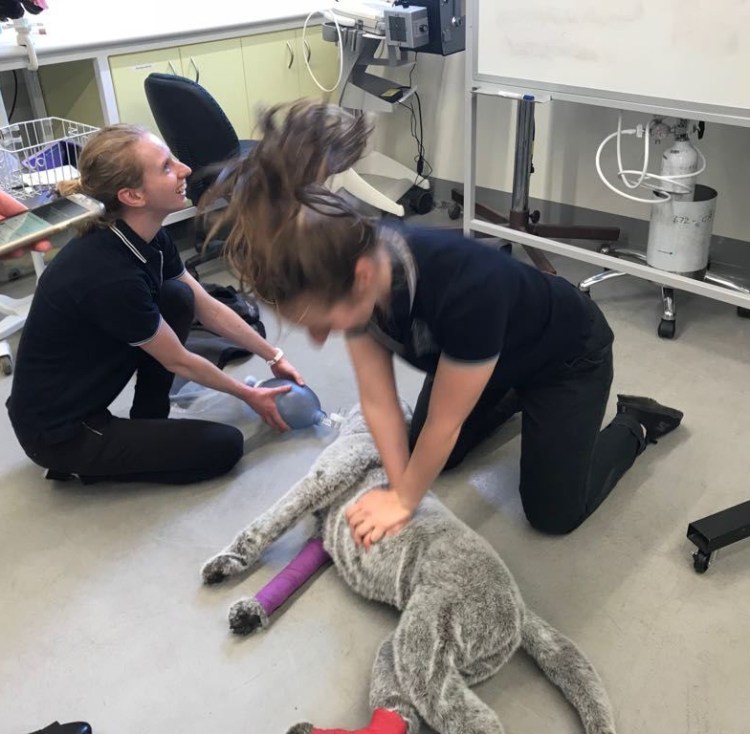
The second component of our diagnostics rotation was radiology and imaging. We started with the musculoskeletal system, and then thorax and abdomen. We were given multiple x-ray films from cats, dogs, and horses. We had normal x-rays to look at as well.  There were several radiology cases to work through and then we had to present a couple cases and our findings to the radiologist. When animals in the hospital needed a x-ray our group would go and assist with positioning the patient on the table and exposing the x-ray. We also looked at the images with the specialists in the radiology room. On the final day of the rotation we had a ‘positioning assessment’ where we had to position a stuffed dog model for different radiographic views.

20180525_134757

My group in the CT room practicing the positioning of ‘Emily’ for her x-rays

I have always enjoyed ultrasounds, so I thought it was very cool when we got some hands on ultrasound practice during this rotation. The first time we got to practice was on the ‘phantom’ which is a specially made box with spheres and wires hidden inside gel that we used the probe to detect. The phantom helps us learn how to manipulate the settings on the ultrasound to better find an image.  The next day we had a LIVE dog to practice on!  We started by finding the kidney in a longitudinal plane and then moving the ultrasound probe to image it in a different view, then moving the probe around the abdomen to look at the spleen, liver, intestines, and bladder. We were supposed to have a 3rd day to practice with the ultrasound and a live patient, but there was no dog available to us. We ended up ultrasounding each other—I am happy to report that I have a gall bladder but possibly only 1 kidney (jk I probably have two kidneys but we couldn’t find the other one).

Anatomic Pathology

Post mortems can be completed on animals as a diagnostic test to help determine the cause of death–especially if it was unable to be determined while the animal was still alive. The first post mortem we did was a congo buffalo from the free range zoo.  Before she died she was walking very weirdly; Her movements looked similar to a disease we see in horses called ‘Stringhalt’. She was immobilized with Etorphine prior to euthanasia. Etorphine is a drug that is very dangerous to humans and can be absorbed through skin and cause immediate cardiac arrest (depending on dose). Because of this we had to wear full PPE (personal protective equipment) and be briefed on what symptoms to watch out for in ourselves and our colleagues. We had someone on standby with a reversal drug. As well, we marked off the leg that the buffalo was darted in & removed that leg prior to starting our necropsy.

congo buffalo

A Congo Buffalo! (No, I also had no idea what they looked like before we saw her)

The second post mortem we performed was a dog that had died due to a dog attack. It was very interesting to see the full extent of the damage from the bite wounds that were not visible externally. The damage likely wouldn’t have been able to be identified with any imaging modalities either.  This experience will influence how I treat future trauma and dog attack cases because I am now better able to understand how many ‘hidden problems’ these patients have.

Our 3rd and most exciting post mortem was a whale! My group was very lucky to be involved in this post mortem.  The whale was stranded in shallow water and was unable to be resuscitated.  Our group and our pathology professors took a little field trip down to the zoo to perform the necropsy at their facilities. We wore full PPE again to protect ourselves against diseases that we might pick up from a marine mammal. Our team was organized—one person was in charge of measurements, ensuring all appropriate samples were collected & organized, others were in charge of examining different body systems. We discovered a massive gastric impaction with sea grass. When we opened one of the stomach compartments there was a large sheet of plastic as well. After doing some research for our post mortem report, we learned that this species of whale eats squid, not sea grass. We suspect that after eating the plastic she may been more inclined to eat an abnormal diet and started eating the sea grass because she had abdominal pain. Unfortunately, she was also pregnant.

32512068_10155161941376394_6060239459276816384_n

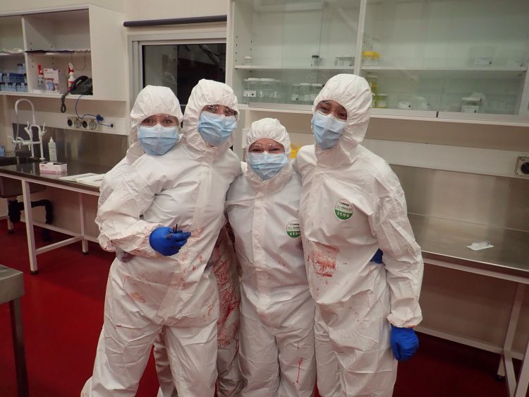
Hanging out in our PPE suits! 

My favourite part of this rotation was the post mortems, especially because we got to work with some very interesting species. However, I’m excited to move on to the next one and get more action in my day!


The First Cat and Dog Rotation!

I feel like there is a significant lack of cat and dog jokes around to make into a quirky title for this blog…

After my group finished our equine rotations we needed to switch our brains into ‘smallies’ mode because we had 3 weeks of Emergency and Critical Care (ECC) and Small Animal Medicine rotation ahead of us.

The Intensive Care Unit (ICU) room is located in the middle of the university hospital and all the most sick or critical patients get transferred there. Often the team is so busy they don’t leave the room and have no idea whats going on in the other departments!  We either worked day shifts or evening shifts–which were the ones I preferred.  This meant that after normal day time hours most of the other hospital departments had gone home–so if a patient needed a special procedure or diagnostic test we did it ourselves instead of transferring to another department.

Again, as a student we were meant to take a case and be “in-charge” of the treatment and monitoring of that animal while it was in hospital. If an animal presented to the hospital it was called over the loudspeaker and we went to the reception area to triage the patient and consult with the owners.

The first patient I saw ended up being the most complicated case we had all week! The cat was initially suspected to have hepatopathy (a fancy way of saying liver disease) and renal (kidney) disease. After further treatment and diagnostics we discovered both an intussusception and a gastric foreign body with bi-cavitary effusions, hypothermia, hypovolemia, and hypotension…which is a real fancy way of saying that this cat was very very sick!  Each day we had rounds where we could sit outside in the gardens and discuss emergency topics. This patient was a great patient to initiate discussions on identification of shock and management of critically ill patients.

On our next shift we had a blocked cat, which is something I had seen a lot of at my previous job! It is not an un-common problem in male cats.  My housemate went to a conference last year and listened to a lecture about sacroiliac blocks (epidurals) for cats during this procedure. We use epidurals quite commonly in cattle but less so in our small animal patients. One night we were lucky enough to have a cadaver cat to practice epidurals on as well as other emergency skills like jugular catheters, tracheotomies, thoracocentesis, and urethral catheterization.

Another evening, there must have been something in the water in Werribee because we had multiple patients present after they feasted on the laundry room products!! One dog ate everything in site—detergent, soap, wine, bleach, smashed glass, etc. One cat ate lily laundry detergent and lilies are particularity toxic to cats!!

In Australia it is quite common to see snake bite cases! The university is currently working on SnakeMap which is this cool project that collects GPS coordinates of where snake bites occur which will help veterinarians manage the cases more efficiently. We had a few patients present to us in different stages of the course of the disease. It was fun being involved in these cases and I will miss them when I practice in Canada one day.

In the true spirit of emergency we had a couple cases which were rushed directly to the ICU room.  My group stood back in awe and watched the well-oiled ECC team perform CPR on a ferret who was involved in a dog attack.  Within minutes we had experts from the anesthesia team and the exotic specialist vet at the ICU room to assist with the uncommon patient.  Another patient in the ICU needed a pericardiocentesis preformed.   This is a procedure where the vet will stick a special needle through the body wall into the sac surrounding the heart (without poking the heart) to remove fluid—often blood–which is preventing the patient from breathing and pumping blood around their body properly.  This is a really cool technique because it is both diagnostic & therapeutic. This means that by performing a pericardiocentesis we can often get a diagnosis of the disease we are dealing with and we can also TREAT that condition at the same time.

WhatsApp Image 2018-04-22 at 10.06.35 PM

Practicing CPR on stuffed animals during our tutorial. But with that hair-do…. I’m not sure if its CPR or headbanging 😛

My group then transitioned into the Small Animal Medicine Department for a slower paced and more detailed rotation. We had tutorials on nutrition for cats and dogs and discussed diets required for different medical conditions and how to best formulate that into a treatment management plan. We also talked about antibiotics and prudent use.  Lastly, we had a CPR tutorial where we practiced our technique on life-sized stuffed animal patients.  We had a ton of fun practicing because we searched up YouTube and played “Stayin’ Alive” on loud to help us keep on time (compressions should be done at a tempo of 120 beats/min)!

I was in charge of another critically ill patient this week that was transferred back and forth from Medicine, ECC, and Surgery. This cat initially presented with blood in his urine. However, he was very anemic as well. Over the course of the week he ended up needing at least 2 blood transfusions. At one point we were considering giving him dog blood (xenotransfusion) because we didn’t have any feline donors.  Personally, I didn’t know that you could even do that without an animal dying and was completely mind blown! The cat continued to get sicker during the week as we tried to figure out an explanation for his many problems. We performed x-rays, ultrasounds, multiple blood tests, and even an exploratory laparotomy (surgery).  This cat had a very guarded prognosis and I was amazed and delighted at the end of the week when he was pacing around the ward and meowing at me for more food in his bowl (and not through his stomach tube!).

Wednesday was cardiology day! We shadowed the specialist cardiologist that comes to the university. My group sat in on all his morning appointments to watch echocardiograms (an ultrasound of the heart) and ECGs. I had a patient that had come in earlier in the week for something unrelated and when I did my physical exam I heard an abnormally low heart rate, a few tests later and an appointment booked in with the cardiologist and we had diagnosed her with a serious heart condition requiring surgical implantation of a pacemaker.

20180313_114208

Sometimes you need several ECG pages to read in order to diagnose the heart condition your patient has…

And on Thursday we had extra training with the specialist oncologist. She was really lovely and spent some time helping us to better feel lymph nodes in dogs and cats. This is something that I have always struggled to find in normal patients who do not have enlarged nodes.  Several of the patients I saw this week were cancer patients in various stages of diagnosis, staging, and treatment.  I now have a better understanding of chemotherapy drugs, what to do when you think you may have found cancer in a patient, how to treat and diagnose cancer.

These 3 weeks provided tons of opportunities for reviewing multiple different diseases and we had a lovely time in the hospital!

The Mane Event

The MANE event! Get it? ha! (It’s probably too late at night to be writing for the public eye…)

After a week “off” for research my group headed back to the equine hospital for our External Equine Rotation. This rotation was meant to be like a ‘general practice horse vet’ but we actually only spent one day driving around to farms to look at horses.

On the first day of the week we used a teaching horse from the university herd and did a practice ‘Pre-Purchase Exam’ on her. This is a special type of physical exam that veterinarians can do on an animal (typically horses or breeding animals) if someone is interested in buying it. There can be a lot of legal implications surrounding these exams– disclosure of medical information, high value of animals, suspected performance status. As a veterinarian you need to know the full extent of your role in this situation. During our exam of the teaching horse we detected some lameness so we took some radiographs of her leg. I have taken many x-rays on cats and dogs but never on horses. It is very difficult to know exactly how to position the horse for x-rays and where to position the machine in order to get the best pictures. This is something I will likely need a lot more practice with if I end up seeing some horses in practice.

Horses have this weird anatomical structure called ‘guttural pouches’ inside their heads. They are a common site of infection in horses so it is important to examine them.   I got a chance to practice driving the scope again. I really like this—its the only “video game” I enjoy.

On Wednesday, my wish came true! The main event! I finally got to see a colic surgery, and not just one, but two!! The horses went into surgery right after each other. I was able to stay on the “dirty side” of the surgery (I didn’t scrub in and work on the “sterile side”) and help out with an impaction colic.  This involves getting rid of all the excessive food material in the horse’s gut that is unable to move through. This was so cool! The second colic surgery was a different kind of colic—this horse had a twist in his intestines.  Can you imagine how painful it would be if your intestines twisted up on themselves? I am so glad that I finally got to see colic surgeries before I finished my equine rotations and be involved in helping these animals recover!

29340143_10155930484875306_1177722582804201472_o

This is just before the surgeons will open the gut to remove the impacted feed material

We worked with another teaching horse from the university herd and practiced placing bandages on his limbs. I much prefer practicing on live animals instead of models or cadaver legs–it is a much more real experience and there is a lot of factors that you learn to deal with i.e: windy day, muddy feet, how to pick up a foot on a horse that doesn’t like his feet being picked up, etc.

20180222_115135

I know this isn’t a limb, but can we all just take a moment to appreciate how great this vet wrap is?!

We also practiced nasogastric tubing (NGT)  horses. This is a really common procedure veterinarians can do to provide a horse with fluids, medications, or decompress a stomach during colic!  It is important to make sure that you put the tube in the stomach of the horse and not the lungs! Quite often we pour fluids down the NGT and we really don’t want to be pouring fluid into the horses lungs.  The way a horse’s larynx & pharynx is placed inside it’s head means that by flexing a horse’s neck downwards while we insert the tube (through the nose) the horse will swallow the tube into the esophagus (where we want it to go!).  You can smell stomach smells from the tube (surprisingly not as bad as you would think…) and hear the stomach bubbling away (a fancy medical version of the game ‘telephone’).

Everyone looks forward to the ambulatory day of this rotation. We drove out to meet one of our wackiest professors for a day of horse vetting in the field! It was a very relaxed day, seeing and chatting about a variety of patients. None of the patients were very sick and it was a pleasant day. We preformed a Caslick procedure, a mini-neurological exam on a mini horse, did some more guttural pouch scoping, and had a lot more fun! Our lunch on this day was legendary! An extremely lovely family made us a feast and I experienced the best date scones I’ve ever eaten…

29258448_10155930482990306_7082194423047520256_o

Me and two of my group mates with our horse vet professor! There was a huge sunflower field just outside one of our appointments.

On the last day of the week we gave a presentation that we had prepared and spoke about our ambulatory cases. Our last practical class this week was on a life-size model horse. We practiced rectal palpation–it is SO important to know the anatomy of a horse (or cow) so that you know what you are feeling (because you can’t see what you are feeling). You can make a lot of diagnoses this way, so the practice was appreciated!

20180222_084652.jpg

My group isn’t going to leave any rotation without some great baking!


Thanks again to the equine team for a wonderful week! We had a blast 🙂

Hay gurl

My trip home over Christmas break was so refreshing. It was just what I needed before heading back to Australia for the final year of my degree. The day I landed back in Melbourne it was 39C. It was a bit of a shock to the system…and naturally being me–I couldn’t wait to get to the beach.

20180128_202224

How is this only 20 mins from my house?

The next morning we started our Equine Internal Rotation at the horse hospital on campus.  This was the rotation that everyone said was difficult, you just have to get through it, and people cry every year. I’m not sure if I took the same rotation or not… but my group had the best time! The whole class is divided up into groups and we spend the  year with those same people as we go through our university rotations.

Whenever a new patient would be admitted to the hospital or come in for a day procedure one of us students had to claim the patient and we would work with that case for the duration of the hospital stay. This also meant that we had to know all the diagnostic tests, findings, and treatments that were run on the patient–we presented everything at the morning and afternoon rounds.

My first case was very interesting. The foal had been admitted to the hospital the day previously with difficulty breathing. It had a tracheostomy tube put in his trachea so he could breath. When I first came in the foal started showing some neurological signs so we decided to investigate further. X-rays showed us that the foal had actually broken it’s neck. This foal needed to be euthanized. I did it; I place very high value on these experiences during vet school.

We call something a ‘textbook case’ when it presents with all the exact signs and symptoms of a disease that you would read about in a textbook. We had a textbook ‘Recurrent Airway Obstruction’ (RAO) case.  It basically means the horse has allergies. It is an important and fairly common equine disease and something that I should likely know well for my board exam! I was pleased that this case came in during my rotation so I could see it in real life and not just read about it. We did a test called a bronchoalveolar lavage (BAL) on the horse which includes putting fluid into the horse’s lungs and then sucking it back up to get a sample of the cells present. Depending on what cells we find this can tell us information about the disease the horse has.

Last year I volunteered in the equine hospital as a 3rd year student. One of the patients that was very memorable came back in for a bit of a check up. This horse is a particularly interesting case because he has a collapsed trachea. This is typically something we see more commonly in dogs and definitely not in horses! His collapsed trachea has already been repaired and he is living comfortably with 2 stents in it to make sure it stays open and he can breathe!

Over the course of the 2 weeks we had a couple of very sick patients–one of them was in isolation. This means we had to start super early, put on all our protective wear and go into his isolation stall to check on him.

Snapchat-1695992760

Luckily there is a cafe in town that opens at 5am. We head there for some real caffeine. 

One of the things I really wanted to see during this rotation was surgery. I specifically was interested in seeing a colic surgery–unfortunately that didn’t happen. But I did get to watch an extremely beautiful Clydesdale undergo an impressive surgical procedure. She had bilateral hock arthroscopy procedures and an ocular cyst removal done. Horse surgery to me is so much more impressive than surgery on cats and dogs. Horses need to be anesthetized and then picked up on a huge winch/crane system so that they can be placed on the surgical table. It can be dangerous to anesthetize horses because of their large size—it causes problems with their lungs! When we first knocked this horse out there was about 13 people running around who all had jobs to prepare the horse for surgery—putting in a urinary catheter, preparing, and scrubbing the surgical sites, applying monitoring equipment, hooking up anesthesia tubes and drugs, positioning the horse, etc etc. Usually I love jumping in and getting involved but this was one time I was happy to be a fly on the wall and just observe all the action around me! It felt like a dream!

Orthopedic work is a big deal in horse medicine. We saw a lot of lame horses during this rotation.  No, a ‘lame horse’ is not a horse we don’t like, it means a horse that is painful in its legs or feet and is not walking normally. This means we watch the horse walk, trot, canter, etc. We check to see if the horse is more lame on a hard surface or a soft surface or while turning or at different speeds. Using a variety of techniques and tests we try to figure out exactly where in the leg the horse is painful! Nerve blocks are a common test we do; we inject a little bit of anesthetic into specific nerves in the leg. This means the horse loses sensation to the area of the leg that that nerve goes to. If we ask the horse to trot and the lameness is gone–then we have a better idea of the location in the leg that the horse is painful!  We can also use x-ray, ultrasound, nuclear scintigraphy, and MRI to help.

Horses are great at injuring themselves! They are impressive and beautiful animals but they are amazing at getting into tricky situations. My next patient was a horse who had been kicked. The wound on his leg went right into the joint space. This can be very dangerous if bacteria are able to grow and then spread around the body. Using a small needle you can stick it directly into the joint and take a sample of his joint fluid. We also flushed out the joint with saline and then left some liquid antibiotics in there before bandaging it all up! We monitored him closely to make sure the infection was healing and not spreading.


A couple of my group’s favorite patients were there for almost the whole 2 weeks of our rotation. One of them was an older teaching horse with a medley of problems but the sweetest personality! We made sure to spend a few extra minutes with her every day. The other horse we loved was a very small stallion who presented with only one eye!  He was very sweet. During his physical exam every day he let me hug him while my partner did the exam.

Snapchat-781925505_LI (2)

A very scientific sample of the discharge taken from the eyeless horse’s socket. We needed to make sure it wasn’t infected. 

Most days we were working 11 or 12 hour days. One day we managed to actually get out of the horse hospital at a decent time and went to the beach right afterwards because it was a nice day. Just as we were getting ready for bed my cell phone rang and we were out the door and back to the hospital for a colicking horse!  Colic is a confusing term–it basically means anything that can give a horse abdominal pain.  An initial colic work up is providing pain medication, rectal palpation (to feel the abdominal organs), checking vital signs, nasogastric tubing, and abdominal ultrasound.  We did all of these tests because we were trying to figure out if the horse needed to go to surgery or if we could treat him medically.  The horse was quite sick but his clinical signs weren’t making sense with any of the more common causes of colic. We decided it was probably best to monitor the horse over night and reassess him the next morning.  During our exam the next day we noticed that he hadn’t urinated since arriving at the hospital and being on IV fluids over night.  I passed a urinary catheter as far as I could up his urethra–until I hit a blockage. Then we put a scope (which is a tiny camera) up his urethra until we saw a stone!! That’s why he couldn’t pee. We did something called a ‘belly tap’ which where we insert a needle into the horse’s abdomen to get a sample of fluid—we discovered that it was urine! This horse had a very rare condition. A stone (we call them uroliths) had formed in his urine and traveled down his urethra until it got stuck and could not move. The horse could not pee and this kept going on until his bladder burst and his abdomen filled up with urine (uroabdomen). Unfortunately, this is very unusual in an adult horse and this horse likely needed at least 2 surgeries to have a chance of recovering—his owner elected to euthanize him. We did the post mortem the next day and the pathology we found definitely confirmed that that was the right decision for this horse.

The next evening we got a referral for a very sick pregnant mare coming in. My whole group decided to stay after hours even though we weren’t all on call.  This horse had hepatic lipidosis –which is a liver disease and was very sick. We were concerned that she was going to abort her foal or go into labor much too early. We decided that she needed to be induced to foal. We prepared all our equipment and debriefed for CPR on the foal.  We were all so tired from being on call and working long days–none of us had food for dinner. We made a call to a kebab shop for sustenance.

We spent a lot of time with that mare and didn’t leave until close to midnight. We had case presentations to give the next day—and we weren’t finished preparing yet! Me and my partner picked the uroabdomen horse to present on because it is such an interesting case! We got home, and finished our presentation and made cupcakes for our last day as a thank you to the equine hospital team! And yes, we modeled them after our patients….

20180209_084539

The cupcakes were modeled after our favourite teaching horse!

Extra big thank you to all the vets, residents, and nurses who put in an effort to teach us, answer our questions, and help us along these past two weeks!